Application
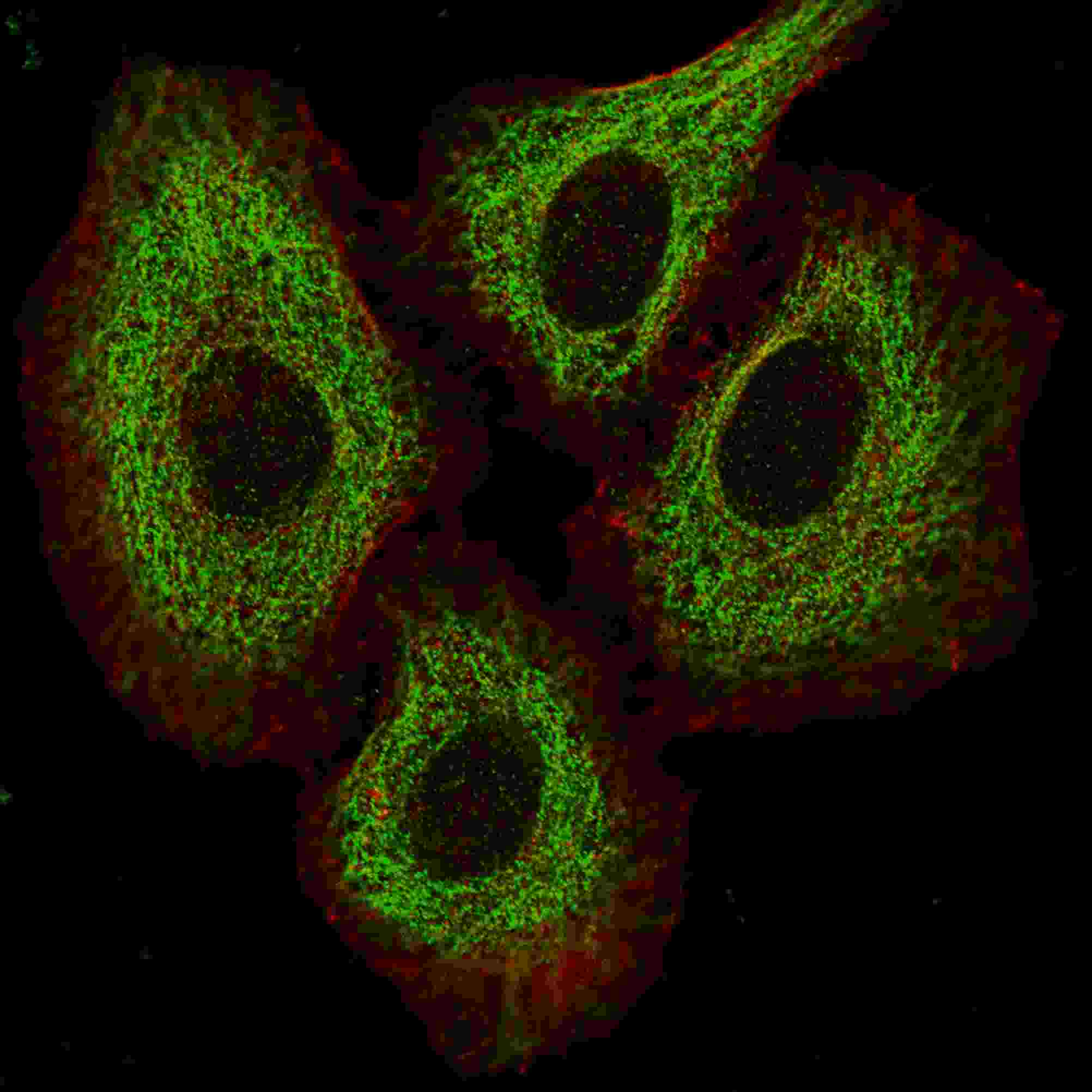
Fluorescent image of HeLa cells stained with TSC2 antibody. HeLa cells were fixed with 4% PFA (20 min), permeabilized with Triton X-100 (0.2%, 30 min). Cells were then incubated with AM1919b TSC2 primary antibody (1:200, 2 h at room temperature). For secondary antibody, Alexa Fluor® 488 conjugated donkey anti-mouse antibody (green) was used (1:1000, 1h). Cytoplasmic actin was counterstained with Alexa Fluor® 555 (red) conjugated Phalloidin (5.25 μM, 25 min). Pictures were taken on a Biorevo microscope (BZ-900, Keyence). Note the highly specific localization of the TSC2 mainly to the cytoplasm, supported by Human Protein Atlas Data (http://www.proteinatlas.org/ENSG00000103197).Application
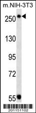
TSC2 Antibody (Cat. #AM1919b) western blot analysis in mouse NIH-3T3 cell line lysates (35ug/lane).This demonstrates the TSC2 antibody detected the TSC2 protein (arrow).| Product Name | TSC2 Antibody |
|---|---|
| Antibody Type | Primary Antibodies |
| Antigen Alias | TSC2; TSC4; Tuberin; Tuberous sclerosis 2 protein |
| Clonality | Monoclonal |
|---|---|
| Isotype | IgG1 |
| Clone Number | 249CT19.1.3 |
| Host Species | Mouse |
| Tested Applications | IFWB |
| IF:1:200 WB |
|
| Species Reactivity | Human |
| Concentration | 1mg/ml |
| Gene Synonyms | TSC4 |
|---|---|
| Alternative Names | TSC2 TSC4 Tuberin Tuberous sclerosis 2 protein |
| Molecular Weight(MW) | 200608 Da |
| Function | In complex with TSC1, inhibits the nutrient-mediated or growth factor-stimulated phosphorylation of S6K1 and EIF4EBP1 by negatively regulating mTORC1 signaling. Acts as a GTPase- activating protein (GAP) for the small GTPase RHEB, a direct activator of the protein kinase activity of mTORC1. Implicated as a tumor suppressor. Involved in microtubule-mediated protein transport, but this seems to be due to unregulated mTOR signaling Stimulates weakly the intrinsic GTPase activity of the Ras-related proteins RAP1A and RAB5 in vitro. Mutations in TSC2 lead to constitutive activation of RAP1A in tumors |
| Tissue Specificity | Liver, brain, heart, lymphocytes, fibroblasts, biliary epithelium, pancreas, skeletal muscle, kidney, lung and placenta |
| Cellular Localization | Cytoplasm. Membrane; Peripheral membrane protein. Note=At steady state found in association with membranes |
| Entrez Gene | 7249 |
|---|---|
| Nucleotide Accession | NP_000539.2 NP_001070651.1 |
Application
Fluorescent image of HeLa cells stained with TSC2 antibody. HeLa cells were fixed with 4% PFA (20 min), permeabilized with Triton X-100 (0.2%, 30 min). Cells were then incubated with AM1919b TSC2 primary antibody (1:200, 2 h at room temperature). For secondary antibody, Alexa Fluor® 488 conjugated donkey anti-mouse antibody (green) was used (1:1000, 1h). Cytoplasmic actin was counterstained with Alexa Fluor® 555 (red) conjugated Phalloidin (5.25 μM, 25 min). Pictures were taken on a Biorevo microscope (BZ-900, Keyence). Note the highly specific localization of the TSC2 mainly to the cytoplasm, supported by Human Protein Atlas Data (http://www.proteinatlas.org/ENSG00000103197).
Application
TSC2 Antibody (Cat. #AM1919b) western blot analysis in mouse NIH-3T3 cell line lysates (35ug/lane).This demonstrates the TSC2 antibody detected the TSC2 protein (arrow).| Application Notes | IF:1:200 WB |
|---|
| Form | Liquid |
|---|---|
| Storage Instructions | For short-term storage, store at 4° C. For long-term storage, aliquot and store at -20ºC or below. Avoid multiple freeze-thaw cycles. |
| Storage Buffer | Purified monoclonal antibody supplied in PBS with 0.09% (W/V) sodium azide. This antibody is purified through a protein G column, eluted with high and low pH buffers and neutralized immediately, followed by dialysis against PBS. |


 Data sheet for OM223757