Application
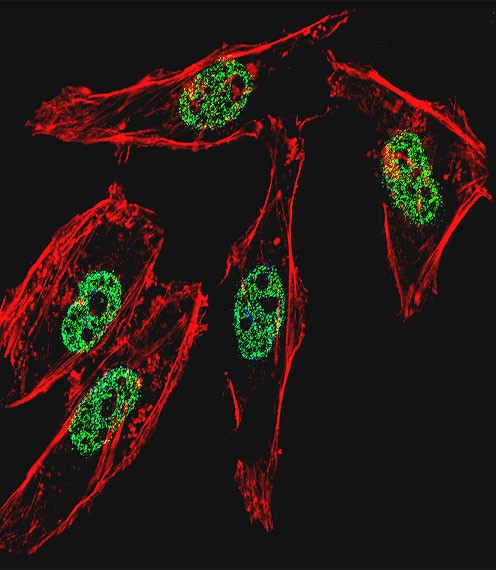
Fluorescent confocal image of Hela cell stained with MyoD1 Antibody . Hela cells were fixed with 4% PFA (20 min), permeabilized with Triton X-100 (0.1%, 10 min), then incubated with MyoD1 primary antibody (1:25). For secondary antibody, Alexa Fluor 488 conjugated donkey anti-rabbit antibody (green) was used (1:400).Cytoplasmic actin was counterstained with Alexa Fluor 555 (red) conjugated PhalloidApplication
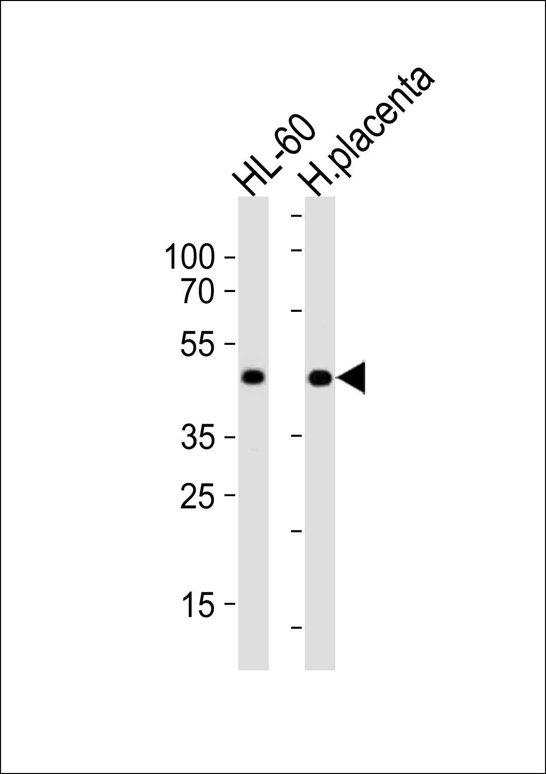
Western blot analysis in Hela cell line and human placenta tissue lysates (35ug/lane).Application
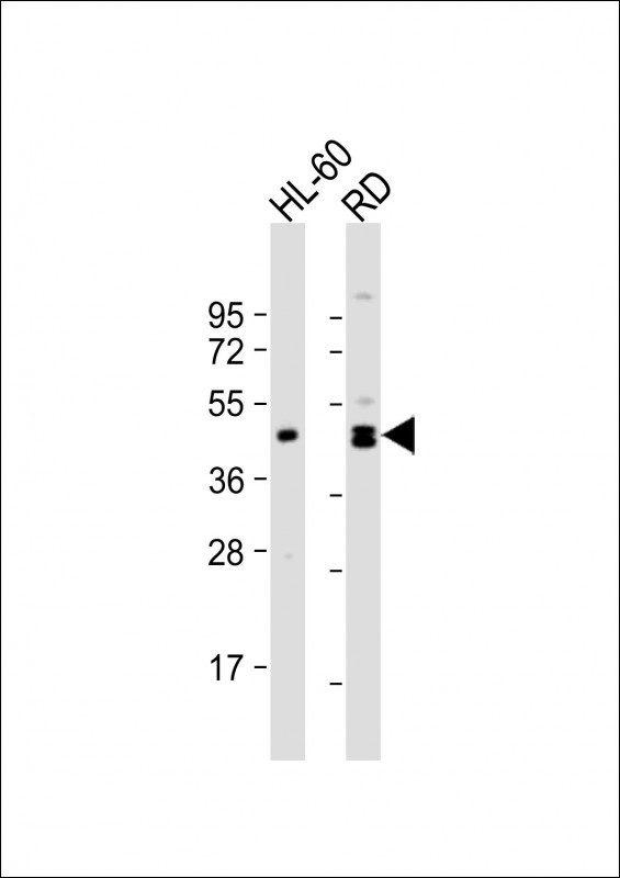
Western Blot at 1:1000 dilution Lane 1: HL-60 whole cell lysate Lane 2: RD whole cell lysate Lysates/proteins at 20 ug per lane.| Product Name | MyoD1 Antibody |
|---|---|
| Antibody Type | Primary Antibodies |
| Product description | This gene encodes a nuclear protein that belongs to the basic helix-loop-helix family of transcription factors and the myogenic factors subfamily. It regulates muscle cell differentiation by inducing cell cycle arrest, a prerequisite for myogenic initiation. The protein is also involved in muscle regeneration. It activates its own transcription which may stabilize commitment to myogenesis.1) Xynos, A., et al. Stem Cells 28(5):965-973(2010) |
| Immunogen | This MyoD1 antibody is generated from rabbits immunized with a KLH conjugated synthetic peptide between 211-240 amino acids from the Central region of human MyoD1. |
| Clonality | Polyclonal |
|---|---|
| Isotype | Ig |
| Host Species | Rabbit |
| Tested Applications | IFWB |
| For WB starting dilution is: 1:1000 For IF starting dilution is: 1:10~50 | |
| Species Reactivity | Human |
| Concentration | 1mg/ml |
| Purification | Affinity purified |
| Gene Symbol | MYOD1 |
|---|---|
| Alternative Names | Myoblast determination protein 1 Class C basic helix-loop-helix protein 1 bHLHc1 Myogenic factor 3 Myf-3 MYOD1 BHLHC1 MYF3 MYOD |
| Molecular Weight(MW) | 35 kDa |
Application
Fluorescent confocal image of Hela cell stained with MyoD1 Antibody . Hela cells were fixed with 4% PFA (20 min), permeabilized with Triton X-100 (0.1%, 10 min), then incubated with MyoD1 primary antibody (1:25). For secondary antibody, Alexa Fluor 488 conjugated donkey anti-rabbit antibody (green) was used (1:400).Cytoplasmic actin was counterstained with Alexa Fluor 555 (red) conjugated Phalloid
Application
Western blot analysis in Hela cell line and human placenta tissue lysates (35ug/lane).
Application
Western Blot at 1:1000 dilution Lane 1: HL-60 whole cell lysate Lane 2: RD whole cell lysate Lysates/proteins at 20 ug per lane.| Application Notes | For WB starting dilution is: 1:1000 For IF starting dilution is: 1:10~50 |
|---|
| Form | Liquid |
|---|---|
| Storage Instructions | Store at 4˚C for three months and -20˚C, stable for up to one year. As with all antibodies care should be taken to avoid repeated freeze thaw cycles. Antibodies should not be exposed to prolonged high temperatures. |
| Storage Buffer | Supplied in PBS with 0.09% (W/V) sodium azide. |


 Data sheet for OM285684