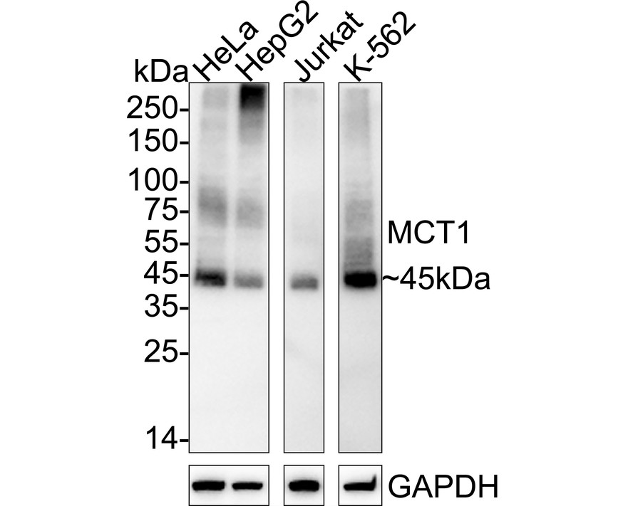
WB
Western blot analysis of MCT1 on different lysates with Rabbit anti-MCT1 antibody at 1/1,000 dilution. Lane 1: HeLa cell lysate, Lane 2: HepG2 cell lysate, Lane 3: Jurkat cell lysate, Lane 4: K-562 cell lysate, Lysates/proteins at 20 µg/Lane. Exposure time: 2 minutes; 4-20% SDS-PAGE gel. Proteins were transferred to a PVDF membrane and blocked with 5% NFDM/TBST for 1 hour at room temperature. The primary antibody at 1/1,000 dilution was used in 5% NFDM/TBST at 4℃ overnight. Goat Anti-Rabbit IgG - HRP Secondary Antibody at 1/50,000 dilution was used for 1 hour at room temperature.IHC
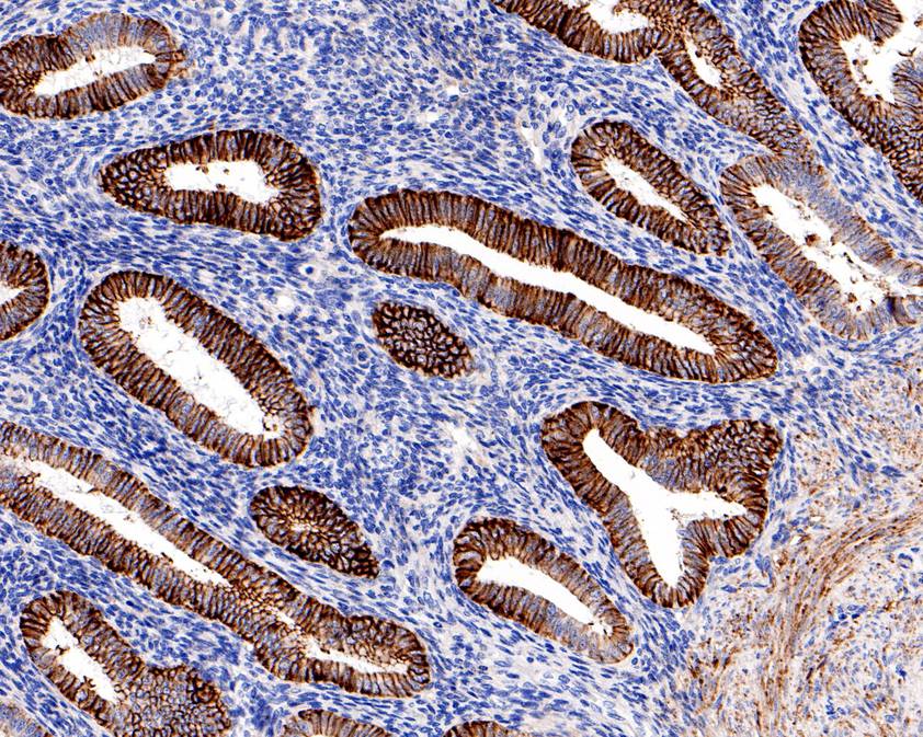
Immunohistochemical analysis of paraffin-embedded human endometrium tissue using anti-MCT1 antibody. The section was pre-treated using heat mediated antigen retrieval with Tris-EDTA buffer (pH 9.0) for 20 minutes.The tissues were blocked in 1% BSA for 30 minutes at room temperature, washed with ddH2O and PBS, and then probed with the primary antibody (1/1,000) for 30 minutes at room temperature. The detection was performed using an HRP conjugated compact polymer system. DAB was used as the chromogen. Tissues were counterstained with hematoxylin and mounted with DPX.ICC/IF
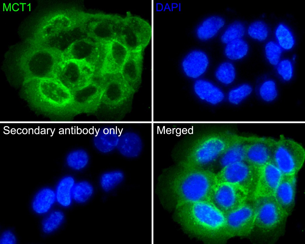
Immunocytochemistry analysis of A431 cells labeling MCT1 with Rabbit anti-MCT1 antibody at 1/200 dilution. Cells were fixed in 4% paraformaldehyde for 10 minutes at 37 ℃, permeabilized with 0.05% Triton X-100 in PBS for 20 minutes, and then blocked with 2% negative goat serum for 30 minutes at room temperature. Cells were then incubated with Rabbit anti-MCT1 antibody at 1/200 dilution in 2% negative goat serum overnight at 4 ℃. Goat Anti-Rabbit IgG H&L (iFluor™ 488) was used as the secondary antibody at 1/1,000 dilution. PBS instead of the primary antibody was used as the secondary antibody only control. Nuclear DNA was labelled in blue with DAPI.FC
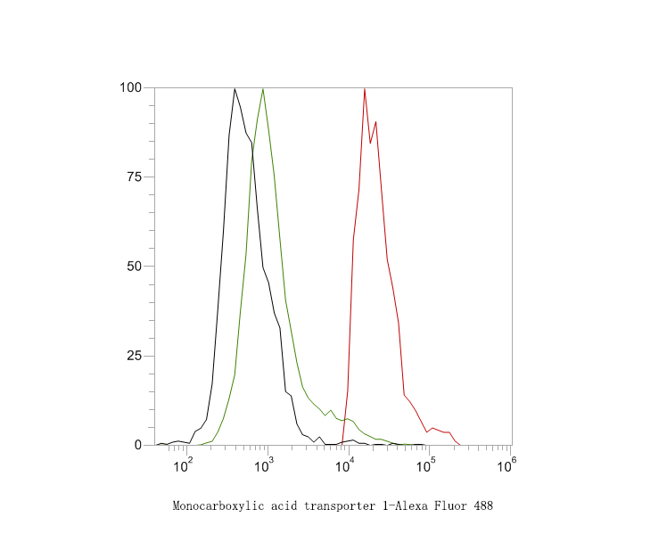
Flow cytometric analysis of MCT1 was done on 293 cells. The cells were stained with the primary antibody (1ug/ml) (red) compared with Rabbit IgG, monoclonal - Isotype Control (green). After incubation of the primary antibody at at +4℃ for 1 hour, the cells were stained with a Alexa Fluor®488 conjugate-Goat anti-Rabbit IgG Secondary antibody at 1/1,000 dilution for 30 min at +4℃ (dark incubation).Unlabelled sample was used as a control (cells without incubation with primary antibody; black).| Product Name | MCT1 Rabbit Polyclonal Antibody |
|---|---|
| Antibody Type | Primary Antibodies |
| Immunogen | Synthetic peptide within human MCT1 aa 450-500. |
| Clonality | Polyclonal |
|---|---|
| Isotype | IgG |
| Host Species | Rabbit |
| Tested Applications | FCICC/IFIHCWB |
| WB:1:1000 IHC:1:200-1:1000 ICC:1:200 FC:1:500-1:1000 |
|
| Species Reactivity | Human |
| Concentration | 1mg/ml |
| Purification | Protein A |
| Gene Symbol | SLC16A1 |
|---|---|
| Gene Synonyms | MCT HHF7 MCT1 MCT1D |
| Gene Full Name | solute carrier family 16 member 1 |
| Gene Summary | The protein encoded by this gene is a proton-linked monocarboxylate transporter that catalyzes the movement of many monocarboxylates, such as lactate and pyruvate, across the plasma membrane. Mutations in this gene are associated with erythrocyte lactate transporter defect. Alternatively spliced transcript variants have been found for this gene.[provided by RefSeq, Oct 2009] |
| Molecular Weight(MW) | 54kDa(Observed band size: 45kDa) |
| Cellular Localization | Cell membrane. |
WB
Western blot analysis of MCT1 on different lysates with Rabbit anti-MCT1 antibody at 1/1,000 dilution. Lane 1: HeLa cell lysate, Lane 2: HepG2 cell lysate, Lane 3: Jurkat cell lysate, Lane 4: K-562 cell lysate, Lysates/proteins at 20 µg/Lane. Exposure time: 2 minutes; 4-20% SDS-PAGE gel. Proteins were transferred to a PVDF membrane and blocked with 5% NFDM/TBST for 1 hour at room temperature. The primary antibody at 1/1,000 dilution was used in 5% NFDM/TBST at 4℃ overnight. Goat Anti-Rabbit IgG - HRP Secondary Antibody at 1/50,000 dilution was used for 1 hour at room temperature.
IHC
Immunohistochemical analysis of paraffin-embedded human endometrium tissue using anti-MCT1 antibody. The section was pre-treated using heat mediated antigen retrieval with Tris-EDTA buffer (pH 9.0) for 20 minutes.The tissues were blocked in 1% BSA for 30 minutes at room temperature, washed with ddH2O and PBS, and then probed with the primary antibody (1/1,000) for 30 minutes at room temperature. The detection was performed using an HRP conjugated compact polymer system. DAB was used as the chromogen. Tissues were counterstained with hematoxylin and mounted with DPX.
ICC/IF
Immunocytochemistry analysis of A431 cells labeling MCT1 with Rabbit anti-MCT1 antibody at 1/200 dilution. Cells were fixed in 4% paraformaldehyde for 10 minutes at 37 ℃, permeabilized with 0.05% Triton X-100 in PBS for 20 minutes, and then blocked with 2% negative goat serum for 30 minutes at room temperature. Cells were then incubated with Rabbit anti-MCT1 antibody at 1/200 dilution in 2% negative goat serum overnight at 4 ℃. Goat Anti-Rabbit IgG H&L (iFluor™ 488) was used as the secondary antibody at 1/1,000 dilution. PBS instead of the primary antibody was used as the secondary antibody only control. Nuclear DNA was labelled in blue with DAPI.
FC
Flow cytometric analysis of MCT1 was done on 293 cells. The cells were stained with the primary antibody (1ug/ml) (red) compared with Rabbit IgG, monoclonal - Isotype Control (green). After incubation of the primary antibody at at +4℃ for 1 hour, the cells were stained with a Alexa Fluor®488 conjugate-Goat anti-Rabbit IgG Secondary antibody at 1/1,000 dilution for 30 min at +4℃ (dark incubation).Unlabelled sample was used as a control (cells without incubation with primary antibody; black).| Application Notes | WB:1:1000 IHC:1:200-1:1000 ICC:1:200 FC:1:500-1:1000 |
|---|
| Form | Liquid |
|---|---|
| Storage Instructions | Store at +4℃ after thawing. Aliquot store at -20℃. Avoid repeated freeze / thaw cycles. |
| Storage Buffer | 1*TBS (pH7.4), 0.2% BSA, 50% Glycerol. Preservative: 0.05% Sodium Azide. |


 Data sheet for OM643500