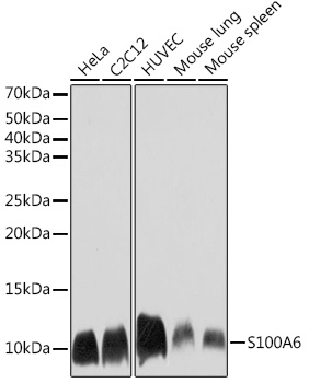
WB
Western blot analysis of various lysates using S100A6 Rabbit mAb at 1:1000 dilution. Secondary antibody: HRP Goat Anti-Rabbit IgG (H+L) at 1:10000 dilution. Lysates/proteins: 25μg per lane. Blocking buffer: 3% nonfat dry milk in TBST. Detection: ECL Basic Kit. Exposure time: 30s.IHC
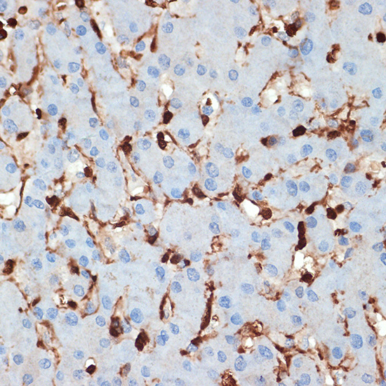
Immunohistochemistry analysis of paraffin embedded human liver using S100A6 Rabbit mAb at dilution of 1:100 (40x lens).Perform microwave antigen retrieval with 10 mM Tris/EDTA buffer pH 9.0 before commencing with IHC staining protocol.ICC/IF
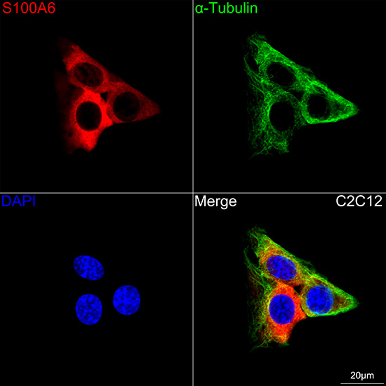
Confocal imaging of C2C12 cells using S100A6 Rabbit mAb (dilution 1:400) followed by a further incubation with Cy3 Goat Anti-Rabbit IgG (H+L) (dilution 1:500) (Red). The cells were counterstained with α-Tubulin Mouse mAb(dilution 1:400) followed by incubation with 488-conjugated Goat Anti-Mouse IgG (H+L) Ab (dilution 1:500)(Green). DAPI was used for nuclear staining (Blue). Objective: 100x.| Product Name | S100A6 Rabbit mAb |
|---|---|
| Antibody Type | Primary Antibodies |
| Immunogen | Recombinant fusion protein containing a sequence corresponding to amino acids 1-90 of human S100A6 (P06703). |
| Clonality | monoclonal |
|---|---|
| Isotype | IgG |
| Host Species | Rabbit |
| Tested Applications | ICC/IFIHCWB |
| WB:1:500-1:12000 IHC:1:50-1:200 ICC/IF:1:100-1:500 |
|
| Species Reactivity | HumanMouseRat |
| Concentration | 1mg/ml |
| Purification | Affinity purified |
| Gene Symbol | S100A6 |
|---|---|
| Gene Synonyms | 2A9 PRA 5B10 CABP CACY S10A6 |
| Gene Full Name | S100 calcium binding protein A6 |
| Gene Summary | The protein encoded by this gene is a member of the S100 family of proteins containing 2 EF-hand calcium-binding motifs. S100 proteins are localized in the cytoplasm and/or nucleus of a wide range of cells, and involved in the regulation of a number of cellular processes such as cell cycle progression and differentiation. S100 genes include at least 13 members which are located as a cluster on chromosome 1q21. This protein may function in stimulation of Ca2+-dependent insulin release, stimulation of prolactin secretion, and exocytosis. Chromosomal rearrangements and altered expression of this gene have been implicated in melanoma. [provided by RefSeq, Jul 2008] |
| Molecular Weight(MW) | 10kDa |
| Cellular Localization | Cell membrane, Cytoplasm, Cytoplasmic side, Nucleus envelope, Peripheral membrane protein, |
WB
Western blot analysis of various lysates using S100A6 Rabbit mAb at 1:1000 dilution. Secondary antibody: HRP Goat Anti-Rabbit IgG (H+L) at 1:10000 dilution. Lysates/proteins: 25μg per lane. Blocking buffer: 3% nonfat dry milk in TBST. Detection: ECL Basic Kit. Exposure time: 30s.
IHC
Immunohistochemistry analysis of paraffin embedded human liver using S100A6 Rabbit mAb at dilution of 1:100 (40x lens).Perform microwave antigen retrieval with 10 mM Tris/EDTA buffer pH 9.0 before commencing with IHC staining protocol.
ICC/IF
Confocal imaging of C2C12 cells using S100A6 Rabbit mAb (dilution 1:400) followed by a further incubation with Cy3 Goat Anti-Rabbit IgG (H+L) (dilution 1:500) (Red). The cells were counterstained with α-Tubulin Mouse mAb(dilution 1:400) followed by incubation with 488-conjugated Goat Anti-Mouse IgG (H+L) Ab (dilution 1:500)(Green). DAPI was used for nuclear staining (Blue). Objective: 100x.| Application Notes | WB:1:500-1:12000 IHC:1:50-1:200 ICC/IF:1:100-1:500 |
|---|
| Form | Liquid |
|---|---|
| Storage Instructions | Store at -20℃. Avoid freeze / thaw cycles. |
| Storage Buffer | Buffer: PBS with 0.05% proclin300, 0.05% BSA, 50% glycerol, pH7.3. |


 Data sheet for OM643933