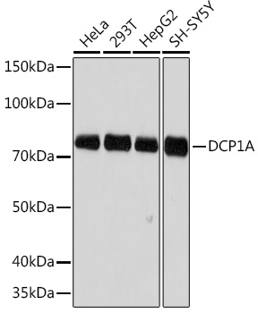

WB
Western blot analysis of various lysates using DCP1A Rabbit mAb at 1:500 dilution. Secondary antibody: HRP-conjugated Goat anti-Rabbit IgG (H+L) at 1:10000 dilution. Lysates/proteins: 25μg per lane. Blocking buffer: 3% nonfat dry milk in TBST. Detection: ECL Basic Kit. Exposure time: 3min.IHC
Immunohistochemistry analysis of paraffin embedded Human esophageal cancer using DCP1A Rabbit mAb at dilution of 1:100 (40x lens). Microwave antigen retrieval performed with 0.01M Tris/EDTA Buffer (pH 9.0) prior to IHC staining.ICC/IF
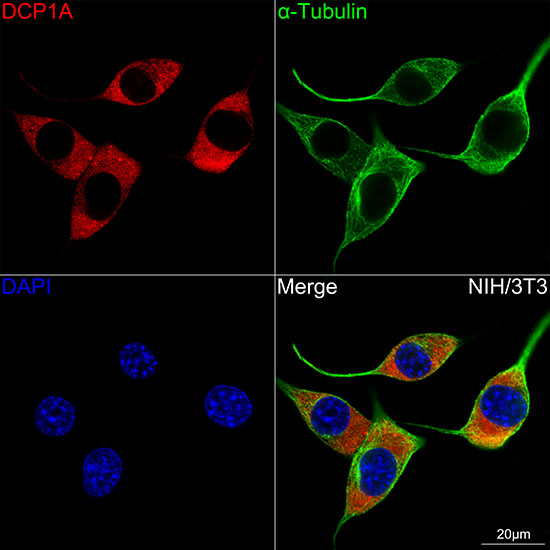
Confocal imaging of NIH/3T3 cells using DCP1A Rabbit mAb (at dilution of 1:100) (Red). The cellsdilution 1:400) (Green). DAPI was used for nuclear staining (blue). Objective: 100x.| Product Name | DCP1A Rabbit mAb |
|---|---|
| Antibody Type | Primary Antibodies |
| Immunogen | A synthetic peptide corresponding to a sequence within amino acids 483-582 of human DCP1A (Q9NPI6). |
| Clonality | monoclonal |
|---|---|
| Isotype | IgG |
| Host Species | Rabbit |
| Tested Applications | ICC/IFIHCWB |
| WB:1:500-1:2000 IHC:1:100-1:1000 ICC/IF:1:50-1:200 |
|
| Species Reactivity | HumanMouse |
| Concentration | 1mg/ml |
| Purification | Affinity purified |
| Gene Symbol | DCP1A |
|---|---|
| Gene Synonyms | SMIF SMAD4IP1 HSA275986 Nbla00360 |
| Gene Full Name | decapping mRNA 1A |
| Gene Summary | Decapping is a key step in general and regulated mRNA decay. The protein encoded by this gene is a decapping enzyme. This protein and another decapping enzyme form a decapping complex, which interacts with the nonsense-mediated decay factor hUpf1 and may be recruited to mRNAs containing premature termination codons. This protein also participates in the TGF-beta signaling pathway. Alternative splicing of this gene results in multiple transcript variants. [provided by RefSeq, Feb 2014] |
| Molecular Weight(MW) | 63kDa(Observed MW 73kDa) |
| Cellular Localization | Cytoplasm, Nucleus, P-body. |
WB
Western blot analysis of various lysates using DCP1A Rabbit mAb at 1:500 dilution. Secondary antibody: HRP-conjugated Goat anti-Rabbit IgG (H+L) at 1:10000 dilution. Lysates/proteins: 25μg per lane. Blocking buffer: 3% nonfat dry milk in TBST. Detection: ECL Basic Kit. Exposure time: 3min.
IHC
Immunohistochemistry analysis of paraffin embedded Human esophageal cancer using DCP1A Rabbit mAb at dilution of 1:100 (40x lens). Microwave antigen retrieval performed with 0.01M Tris/EDTA Buffer (pH 9.0) prior to IHC staining.
ICC/IF
Confocal imaging of NIH/3T3 cells using DCP1A Rabbit mAb (at dilution of 1:100) (Red). The cellsdilution 1:400) (Green). DAPI was used for nuclear staining (blue). Objective: 100x.| Application Notes | WB:1:500-1:2000 IHC:1:100-1:1000 ICC/IF:1:50-1:200 |
|---|
| Form | Liquid |
|---|---|
| Storage Instructions | Store at -20℃. Avoid freeze / thaw cycles. |
| Storage Buffer | Buffer: PBS with 0.05% proclin300, 0.05% BSA, 50% glycerol, pH7.3. |


 Data sheet for OM643989