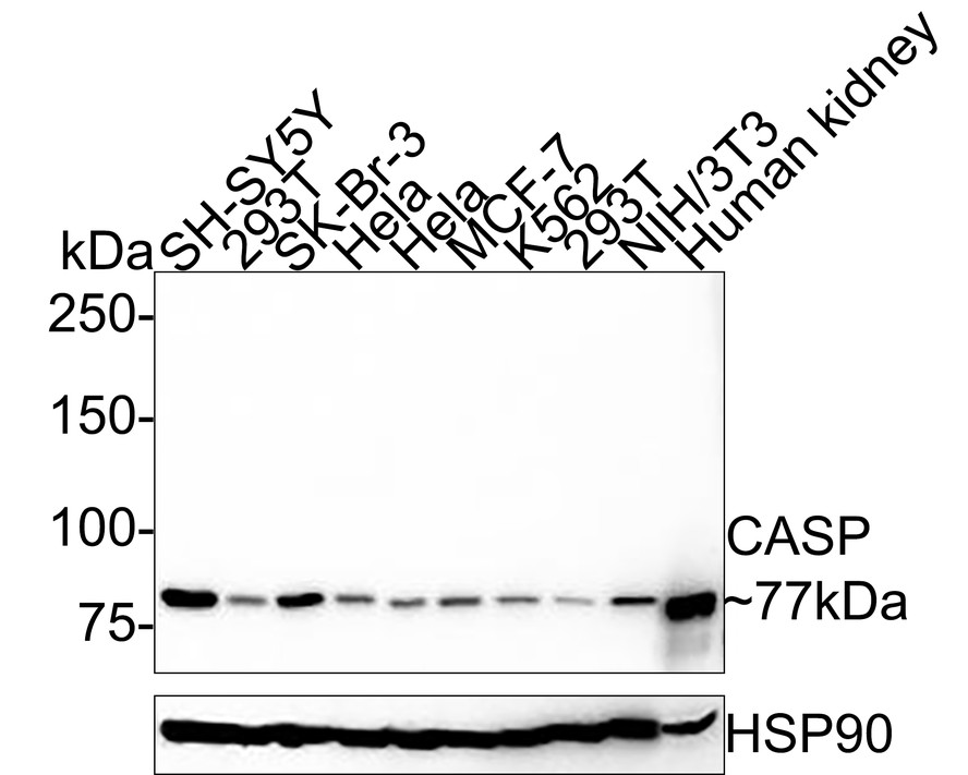
WB
Western blot analysis of Protein CASP on different lysates with Mouse anti-Protein CASP antibody at 1/5,000 dilution. Lane 1: SH-SY5Y cell lysate (20 µg/Lane), Lane 2: 293T cell lysate (20 µg/Lane), Lane 3: SK-Br-3 cell lysate (20 µg/Lane), Lane 4: Hela cell lysate (20 µg/Lane), Lane 5: Hela cell lysate (20 µg/Lane), Lane 6: MCF-7 cell lysate (20 µg/Lane), Lane 7: K562 cell lysate (20 µg/Lane), Lane 8: 293T cell lysate (20 µg/Lane), Lane 9: NIH/3T3 cell lysate (20 µg/Lane), Lane 10: Human kidney tissue lysate (40 µg/Lane), Exposure time: 19 seconds; 6% SDS-PAGE gel. Proteins were transferred to a PVDF membrane and blocked with 5% NFDM/TBST for 1 hour at room temperature. The primary antibody at 1/5,000 dilution was used in 5% NFDM/TBST at room temperature for 2 hours. Goat Anti-Mouse IgG - HRP Secondary Antibody at 1:150,000 dilution was used for 1 hour at room temperature.IHC
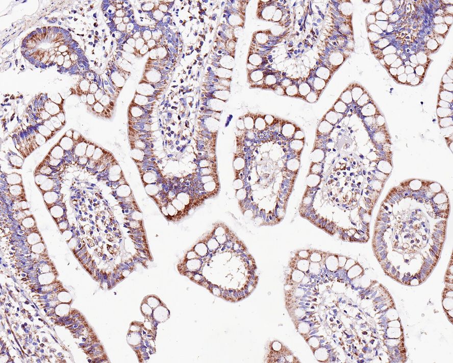
Immunohistochemical analysis of paraffin-embedded human small intestine tissue with Mouse anti-Protein CASP antibody at 1ug/mL dilution. The section was pre-treated using heat mediated antigen retrieval with sodium citrate buffer (pH 6.0) for 2 minutes. The tissues were blocked in 1% BSA for 20 minutes at room temperature, washed with ddH2O and PBS, and then probed with the primary antibody at 1ug/mL dilution for 1 hour at room temperature. The detection was performed using an HRP conjugated compact polymer system. DAB was used as the chromogen. Tissues were counterstained with hematoxylin and mounted with DPX.IHC
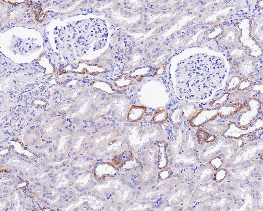
Immunohistochemical analysis of paraffin-embedded human kidney tissue with Mouse anti-Protein CASP antibody at 1ug/mL dilution. The section was pre-treated using heat mediated antigen retrieval with sodium citrate buffer (pH 6.0) for 2 minutes. The tissues were blocked in 1% BSA for 20 minutes at room temperature, washed with ddH2O and PBS, and then probed with the primary antibody at 1ug/mL dilution for 1 hour at room temperature. The detection was performed using an HRP conjugated compact polymer system. DAB was used as the chromogen. Tissues were counterstained with hematoxylin and mounted with DPX.ICC/IF
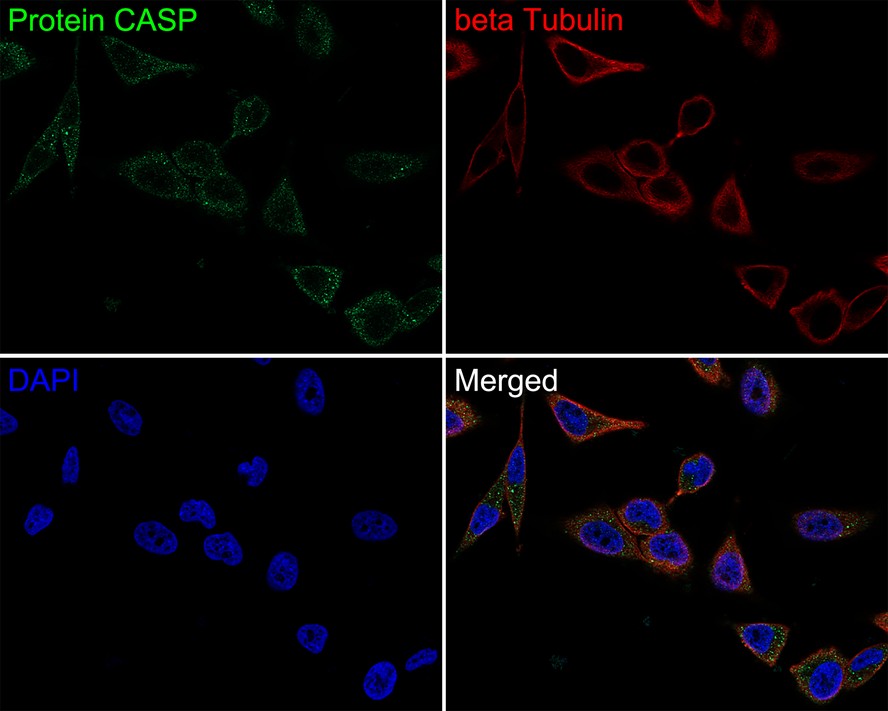
Immunocytochemistry analysis of Hela cells labeling Protein CASP with Mouse anti-Protein CASP antibody at 1ug/mL dilution. Cells were fixed in 4% paraformaldehyde for 30 minutes, permeabilized with 0.05% Triton X-100 in PBS for 20 minutes, and then blocked with 2% negative goat serum for 30 minutes at room temperature. Cells were then incubated with Mouse anti-Protein CASP antibody at 1ug/mL dilution in 2% negative goat serum overnight at 4 ℃. Goat Anti-Mouse IgG H&L (488) was used as the secondary antibody at 1/1,000 dilution. Nuclear DNA was labelled in blue with DAPI. beta Tubulin (red) was stained at 1/100 dilution overnight at +4℃. Goat Anti-Rabbit IgG H&L (594) were used as the secondary antibody at 1/1,000 dilution.| Product Name | Protein CASP Mouse Monoclonal Antibody |
|---|---|
| Antibody Type | Primary Antibodies |
| Immunogen | Recombinant protein within human Protein CASP aa 6-55. |
| Clonality | monoclonal |
|---|---|
| Isotype | IgG1 |
| Host Species | Mouse |
| Tested Applications | ICC/IFIHCWB |
| WB:1:5000 IHC:1:200-1:1000 ICC/IF:1:200-1:1000 |
|
| Species Reactivity | HumanMouseRat |
| Concentration | 1mg/ml |
| Purification | Protein A |
| Gene Symbol | CUX1 |
|---|---|
| Gene Synonyms | CDP CUX p75 CASP CDP1 COY1 Clox GDDI p100 p110 p200 CUTL1 GOLIM6 CDP/Cut Cux/CDP Nbla10317 |
| Gene Full Name | cut like homeobox 1 |
| Gene Summary | The protein encoded by this gene is a member of the homeodomain family of DNA binding proteins. It may regulate gene expression, morphogenesis, and differentiation and it may also play a role in the cell cycle progession. Several alternatively spliced transcript variants encoding different isoforms have been identified.[provided by RefSeq, Feb 2011] |
| Molecular Weight(MW) | 77kDa |
| Cellular Localization | Golgi apparatus membrane. |
WB
Western blot analysis of Protein CASP on different lysates with Mouse anti-Protein CASP antibody at 1/5,000 dilution. Lane 1: SH-SY5Y cell lysate (20 µg/Lane), Lane 2: 293T cell lysate (20 µg/Lane), Lane 3: SK-Br-3 cell lysate (20 µg/Lane), Lane 4: Hela cell lysate (20 µg/Lane), Lane 5: Hela cell lysate (20 µg/Lane), Lane 6: MCF-7 cell lysate (20 µg/Lane), Lane 7: K562 cell lysate (20 µg/Lane), Lane 8: 293T cell lysate (20 µg/Lane), Lane 9: NIH/3T3 cell lysate (20 µg/Lane), Lane 10: Human kidney tissue lysate (40 µg/Lane), Exposure time: 19 seconds; 6% SDS-PAGE gel. Proteins were transferred to a PVDF membrane and blocked with 5% NFDM/TBST for 1 hour at room temperature. The primary antibody at 1/5,000 dilution was used in 5% NFDM/TBST at room temperature for 2 hours. Goat Anti-Mouse IgG - HRP Secondary Antibody at 1:150,000 dilution was used for 1 hour at room temperature.
IHC
Immunohistochemical analysis of paraffin-embedded human small intestine tissue with Mouse anti-Protein CASP antibody at 1ug/mL dilution. The section was pre-treated using heat mediated antigen retrieval with sodium citrate buffer (pH 6.0) for 2 minutes. The tissues were blocked in 1% BSA for 20 minutes at room temperature, washed with ddH2O and PBS, and then probed with the primary antibody at 1ug/mL dilution for 1 hour at room temperature. The detection was performed using an HRP conjugated compact polymer system. DAB was used as the chromogen. Tissues were counterstained with hematoxylin and mounted with DPX.
IHC
Immunohistochemical analysis of paraffin-embedded human kidney tissue with Mouse anti-Protein CASP antibody at 1ug/mL dilution. The section was pre-treated using heat mediated antigen retrieval with sodium citrate buffer (pH 6.0) for 2 minutes. The tissues were blocked in 1% BSA for 20 minutes at room temperature, washed with ddH2O and PBS, and then probed with the primary antibody at 1ug/mL dilution for 1 hour at room temperature. The detection was performed using an HRP conjugated compact polymer system. DAB was used as the chromogen. Tissues were counterstained with hematoxylin and mounted with DPX.
ICC/IF
Immunocytochemistry analysis of Hela cells labeling Protein CASP with Mouse anti-Protein CASP antibody at 1ug/mL dilution. Cells were fixed in 4% paraformaldehyde for 30 minutes, permeabilized with 0.05% Triton X-100 in PBS for 20 minutes, and then blocked with 2% negative goat serum for 30 minutes at room temperature. Cells were then incubated with Mouse anti-Protein CASP antibody at 1ug/mL dilution in 2% negative goat serum overnight at 4 ℃. Goat Anti-Mouse IgG H&L (488) was used as the secondary antibody at 1/1,000 dilution. Nuclear DNA was labelled in blue with DAPI. beta Tubulin (red) was stained at 1/100 dilution overnight at +4℃. Goat Anti-Rabbit IgG H&L (594) were used as the secondary antibody at 1/1,000 dilution.| Application Notes | WB:1:5000 IHC:1:200-1:1000 ICC/IF:1:200-1:1000 |
|---|
| Form | Liquid |
|---|---|
| Storage Instructions | Store at +4℃ after thawing. Aliquot store at -20℃. Avoid repeated freeze / thaw cycles. |
| Storage Buffer | 1*TBS (pH7.4), 0.05% BSA, 40% Glycerol. Preservative: 0.05% Sodium Azide. |


 Data sheet for OM644048