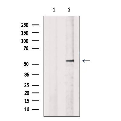
WB
Western blot analysis of extracts from Myeloma cells, using OLFM4 Antibody. The lane on the left was treated with blocking peptide.ICC/IF
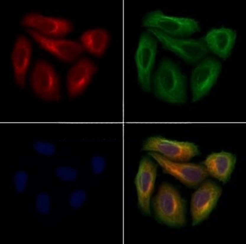
OLFM4 Antibody staining Hela cells by ICC/IF. The samples were fixed with PFA and permeabilized in 0.1% Triton X-100,then blocked in 10% serum for 45 minutes at 25°C. Samples were then incubated with primary Ab(1:200) and mouse anti-beta tubulin Ab(1:200) for 1 hour at 37°C. An AlexaFluor594 conjugated goat anti-rabbit IgG(H+L) Ab(Red) and an AlexaFluor488 conjugated goat anti-mouse IgG(H+L) Ab(Green) were used as the secondary antibody. The nuclear counter stain is DAPI(blue).IHC
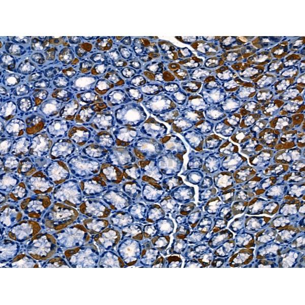
OLFM4 Antibody at 1/100 staining Mouse stomach tissue by IHC-P. The sample was formaldehyde fixed and a heat mediated antigen retrieval step in citrate buffer was performed. The sample was then blocked and incubated with the primary antibody at 4°C overnight. An HRP conjugated anti-Rabbit antibody was used as the secondary antibody.| Product Name | Rabbit polyclonal antibody to OLFM4 |
|---|---|
| Antibody Type | Primary Antibodies |
| Immunogen | A synthesized peptide derived from human OLFM4(Accession Q6UX06), corresponding to amino acid residues N253-Y303. |
| Clonality | polyclonal |
|---|---|
| Isotype | IgG |
| Host Species | Rabbit |
| Tested Applications | ICC/IFIHCWB |
| WB:1:500-1:2000 ICC/IF:1:100-1:500 IHC:1:50-1:200 |
|
| Species Reactivity | HumanMouseRat |
| Concentration | 1mg/ml |
| Purification | Affinity purified |
| Gene Symbol | OLFM4 |
|---|---|
| Gene Synonyms | GC1 OLM4 OlfD pDP4 GW112 hGC-1 hOLfD UNQ362 bA209J19.1 |
| Gene Full Name | olfactomedin 4 |
| Gene Summary | This gene was originally cloned from human myeloblasts and found to be selectively expressed in inflammed colonic epithelium. This gene encodes a member of the olfactomedin family. The encoded protein is an antiapoptotic factor that promotes tumor growth and is an extracellular matrix glycoprotein that facilitates cell adhesion. [provided by RefSeq, Mar 2011] |
| Molecular Weight(MW) | 57kDa |
| Cellular Localization | Secreted>Extracellular space. Mitochondrion. |
WB
Western blot analysis of extracts from Myeloma cells, using OLFM4 Antibody. The lane on the left was treated with blocking peptide.
ICC/IF
OLFM4 Antibody staining Hela cells by ICC/IF. The samples were fixed with PFA and permeabilized in 0.1% Triton X-100,then blocked in 10% serum for 45 minutes at 25°C. Samples were then incubated with primary Ab(1:200) and mouse anti-beta tubulin Ab(1:200) for 1 hour at 37°C. An AlexaFluor594 conjugated goat anti-rabbit IgG(H+L) Ab(Red) and an AlexaFluor488 conjugated goat anti-mouse IgG(H+L) Ab(Green) were used as the secondary antibody. The nuclear counter stain is DAPI(blue).
IHC
OLFM4 Antibody at 1/100 staining Mouse stomach tissue by IHC-P. The sample was formaldehyde fixed and a heat mediated antigen retrieval step in citrate buffer was performed. The sample was then blocked and incubated with the primary antibody at 4°C overnight. An HRP conjugated anti-Rabbit antibody was used as the secondary antibody.| Application Notes | WB:1:500-1:2000 ICC/IF:1:100-1:500 IHC:1:50-1:200 |
|---|
| Form | Liquid |
|---|---|
| Storage Instructions | Store at -20 °C. Stable for 12 months from date of receipt. |
| Storage Buffer | Rabbit IgG in phosphate buffered saline , pH 7.4, 150mM NaCl, 0.02% sodium azide and 50% glycerol. |


 Data sheet for OM644202