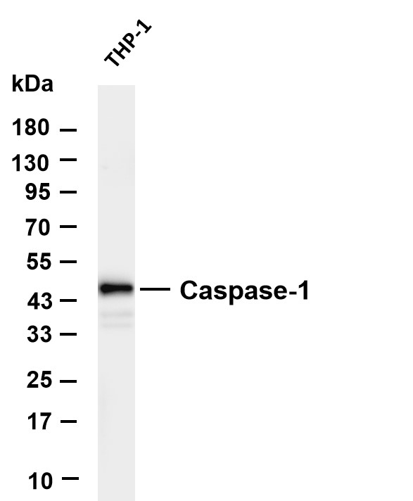
WB
Various whole cell lysates were separated by 4-20% SDS-PAGE, and the membrane was blotted with anti-Caspase-1 antibody. The HRP-conjugated Goat anti-Rabbit IgG(H + L) antibody was used to detect the antibody. Lane 1: THP-1.IHC
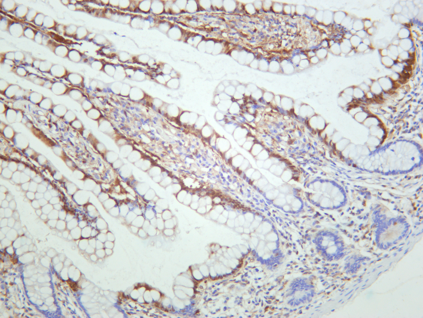
Human small intestine was stained with anti-Caspase-1 rabbit antibody.ICC/IF
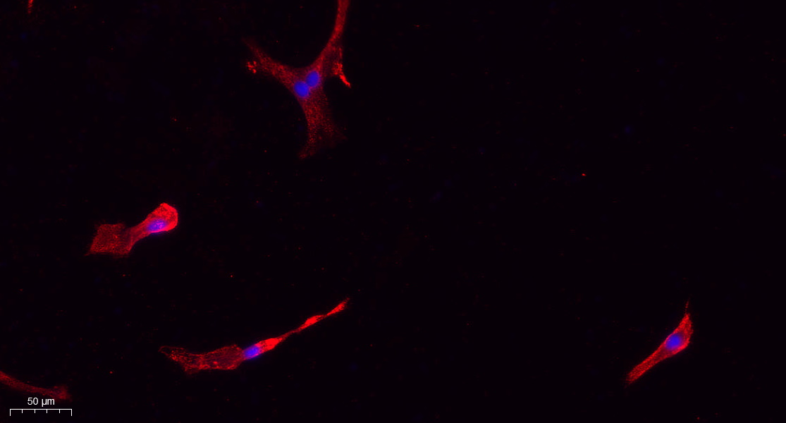
Immunofluorescence analysis of A549. 1,primary Antibody(red) was diluted at 1:200(4°C overnight). 2, Goat Anti Rabbit IgG (H&L) - Alexa Fluor 594 Secondary antibody was diluted at 1:1000(room temperature, 50min).3, Picture B: DAPI(blue) 10min.| Product Name | Caspase-1 Rabbit mAb |
|---|---|
| Antibody Type | Primary Antibodies |
| Clonality | monoclonal |
|---|---|
| Isotype | IgG |
| Host Species | Rabbit |
| Tested Applications | ICC/IFIHCWB |
| WB:1:2000-1:10000 IHC:1:200-1:1000 ICC/IF:1:200-1:1000 |
|
| Species Reactivity | HumanMouseRat |
| Concentration | 1mg/ml |
| Purification | Protein A |
| Gene Symbol | CASP1 |
|---|---|
| Gene Synonyms | ICE P45 IL1BC |
| Gene Full Name | caspase 1 |
| Gene Summary | This gene encodes a protein which is a member of the cysteine-aspartic acid protease (caspase) family. Sequential activation of caspases plays a central role in the execution-phase of cell apoptosis. Caspases exist as inactive proenzymes which undergo proteolytic processing at conserved aspartic residues to produce 2 subunits, large and small, that dimerize to form the active enzyme. This gene was identified by its ability to proteolytically cleave and activate the inactive precursor of interleukin-1, a cytokine involved in the processes such as inflammation, septic shock, and wound healing. This gene has been shown to induce cell apoptosis and may function in various developmental stages. Studies of a similar gene in mouse suggest a role in the pathogenesis of Huntington disease. Alternative splicing results in transcript variants encoding distinct isoforms. [provided by RefSeq, Mar 2012] |
| Molecular Weight(MW) | 45kDa |
| Cellular Localization | Cytoplasm. |
WB
Various whole cell lysates were separated by 4-20% SDS-PAGE, and the membrane was blotted with anti-Caspase-1 antibody. The HRP-conjugated Goat anti-Rabbit IgG(H + L) antibody was used to detect the antibody. Lane 1: THP-1.
IHC
Human small intestine was stained with anti-Caspase-1 rabbit antibody.
ICC/IF
Immunofluorescence analysis of A549. 1,primary Antibody(red) was diluted at 1:200(4°C overnight). 2, Goat Anti Rabbit IgG (H&L) - Alexa Fluor 594 Secondary antibody was diluted at 1:1000(room temperature, 50min).3, Picture B: DAPI(blue) 10min.| Application Notes | WB:1:2000-1:10000 IHC:1:200-1:1000 ICC/IF:1:200-1:1000 |
|---|
| Form | Liquid |
|---|---|
| Storage Instructions | -15°C to -25°C/1 year(Do not lower than -25°C) |
| Storage Buffer | PBS, 50% glycerol, 0.05% Proclin 300, 0.05%BSA |


 Data sheet for OM644212