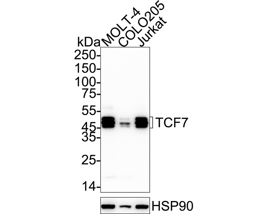
WB
Western blot analysis of TCF7 on different lysates with Rabbit anti-TCF7 antibody at 1/2,000 dilution. Lane 1: MOLT-4 cell lysate, Lane 2: COLO205 cell lysate, Lane 3: Jurkat cell lysate, Lysates/proteins at 20 µg/Lane. Exposure time: 25 seconds; 4-20% SDS-PAGE gel. Proteins were transferred to a PVDF membrane and blocked with 5% NFDM/TBST for 1 hour at room temperature. The primary antibody at 1/2,000 dilution was used in 5% NFDM/TBST at 4℃ overnight. Goat Anti-Rabbit IgG - HRP Secondary Antibody at 1/50,000 dilution was used for 1 hour at room temperature.IHC
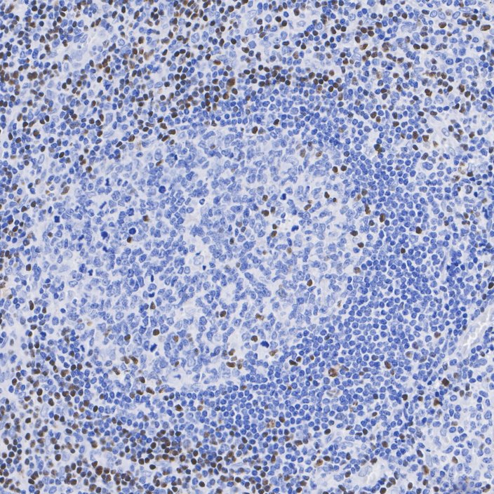
Immunohistochemical analysis of paraffin-embedded human tonsils tissue with Rabbit anti-TCF7 antibody at 1/1,000 dilution. The section was pre-treated using heat mediated antigen retrieval with Tris-EDTA buffer (pH 9.0) for 20 minutes. The tissues were blocked in 1% BSA for 20 minutes at room temperature, washed with ddH2O and PBS, and then probed with the primary antibody at 1/1,000 dilution for 1 hour at room temperature. The detection was performed using an HRP conjugated compact polymer system. DAB was used as the chromogen. Tissues were counterstained with hematoxylin and mounted with DPX.IP
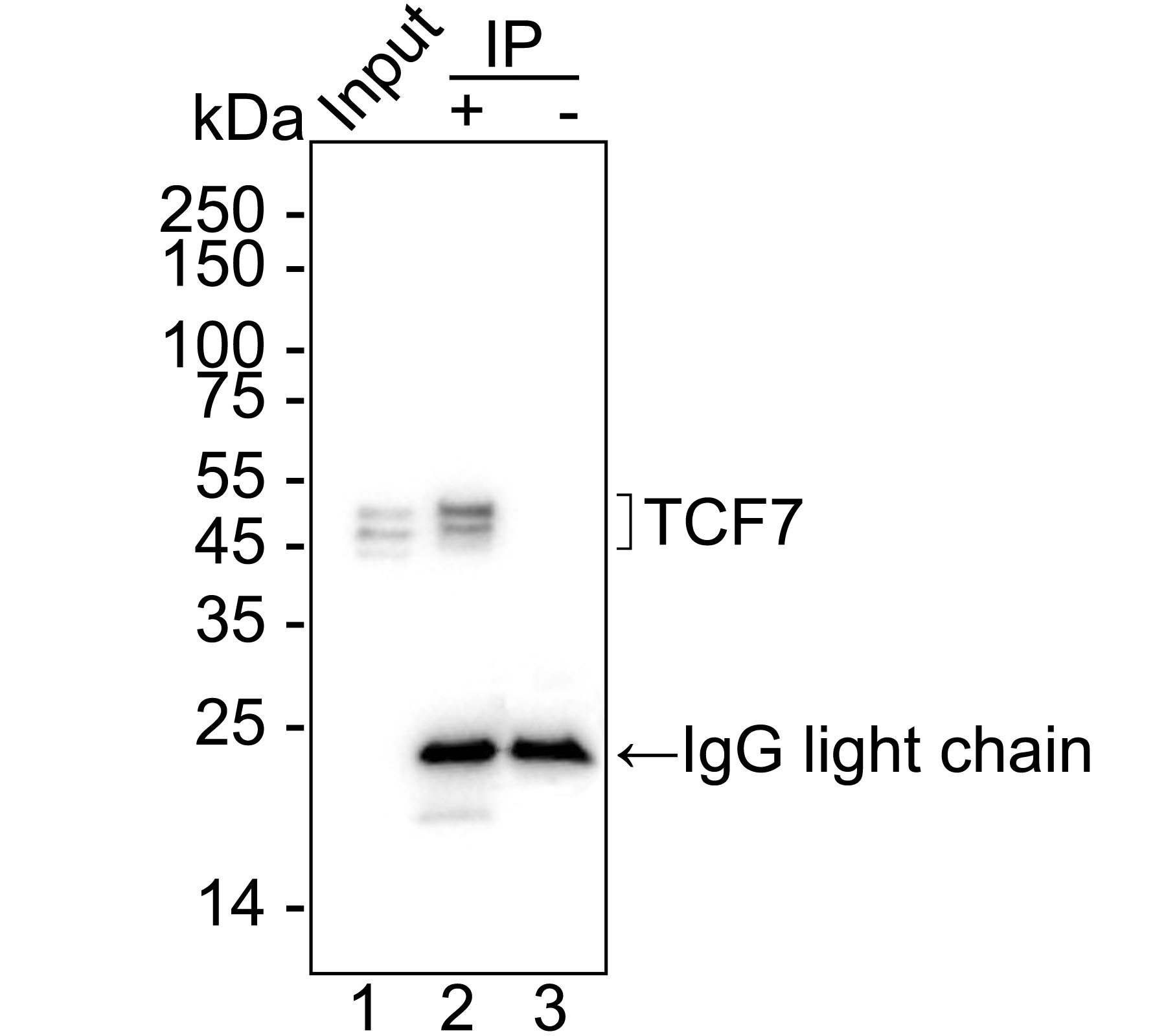
TCF7 was immunoprecipitated from 0.2 mg Jurkat cell lysate withRabbit anti-TCF7 antibody at 2 µg/10 µl beads. Western blot was performed from the immunoprecipitate usingRabbit anti-TCF7 antibody at 1/1,000 dilution. Mouse Anti-Rabbit IgG kappa light chain secondary antibody at 1/5,000 dilution was used for 1 hour at room temperature. Lane 1: Jurkat cell lysate (input), Lane 2: Rabbit anti-TCF7 antibody IP in Jurkat cell lysate, Lane 3: Rabbit IgG instead of Rabbit anti-TCF7 antibody in Jurkat cell lysate, Blocking/Dilution buffer: 5% NFDM/TBST. Exposure time: 1 seconds.| Product Name | TCF7 Recombinant Rabbit Monoclonal Antibody |
|---|---|
| Antibody Type | Primary Antibodies |
| Immunogen | Recombinant protein within Human Netrin 1 aa 505-604 / 604. |
| Clonality | monoclonal |
|---|---|
| Isotype | IgG |
| Host Species | Rabbit |
| Tested Applications | IHCIPWB |
| WB:1:2000 IHC:1:1000 IP:1-2μg/sample |
|
| Species Reactivity | Human |
| Concentration | 1mg/ml |
| Purification | Protein A |
| Gene Symbol | TCF7 |
|---|---|
| Gene Synonyms | TCF-1 |
| Gene Full Name | transcription factor 7 |
| Gene Summary | This gene encodes a member of the T-cell factor/lymphoid enhancer-binding factor family of high mobility group (HMG) box transcriptional activators. This gene is expressed predominantly in T-cells and plays a critical role in natural killer cell and innate lymphoid cell development. The encoded protein forms a complex with beta-catenin and activates transcription through a Wnt/beta-catenin signaling pathway. Mice with a knockout of this gene are viable and fertile, but display a block in T-lymphocyte differentiation. Alternative splicing results in multiple transcript variants. Naturally-occurring isoforms lacking the N-terminal beta-catenin interaction domain may act as dominant negative regulators of Wnt signaling. [provided by RefSeq, Oct 2016] |
| Molecular Weight(MW) | 42kDa(Observed band size: 45-50kDa) |
| Cellular Localization | Nucleus. |
WB
Western blot analysis of TCF7 on different lysates with Rabbit anti-TCF7 antibody at 1/2,000 dilution. Lane 1: MOLT-4 cell lysate, Lane 2: COLO205 cell lysate, Lane 3: Jurkat cell lysate, Lysates/proteins at 20 µg/Lane. Exposure time: 25 seconds; 4-20% SDS-PAGE gel. Proteins were transferred to a PVDF membrane and blocked with 5% NFDM/TBST for 1 hour at room temperature. The primary antibody at 1/2,000 dilution was used in 5% NFDM/TBST at 4℃ overnight. Goat Anti-Rabbit IgG - HRP Secondary Antibody at 1/50,000 dilution was used for 1 hour at room temperature.
IHC
Immunohistochemical analysis of paraffin-embedded human tonsils tissue with Rabbit anti-TCF7 antibody at 1/1,000 dilution. The section was pre-treated using heat mediated antigen retrieval with Tris-EDTA buffer (pH 9.0) for 20 minutes. The tissues were blocked in 1% BSA for 20 minutes at room temperature, washed with ddH2O and PBS, and then probed with the primary antibody at 1/1,000 dilution for 1 hour at room temperature. The detection was performed using an HRP conjugated compact polymer system. DAB was used as the chromogen. Tissues were counterstained with hematoxylin and mounted with DPX.
IP
TCF7 was immunoprecipitated from 0.2 mg Jurkat cell lysate withRabbit anti-TCF7 antibody at 2 µg/10 µl beads. Western blot was performed from the immunoprecipitate usingRabbit anti-TCF7 antibody at 1/1,000 dilution. Mouse Anti-Rabbit IgG kappa light chain secondary antibody at 1/5,000 dilution was used for 1 hour at room temperature. Lane 1: Jurkat cell lysate (input), Lane 2: Rabbit anti-TCF7 antibody IP in Jurkat cell lysate, Lane 3: Rabbit IgG instead of Rabbit anti-TCF7 antibody in Jurkat cell lysate, Blocking/Dilution buffer: 5% NFDM/TBST. Exposure time: 1 seconds.| Application Notes | WB:1:2000 IHC:1:1000 IP:1-2μg/sample |
|---|
| Form | Liquid |
|---|---|
| Storage Instructions | Store at +4℃ after thawing. Aliquot store at -20℃. Avoid repeated freeze / thaw cycles. |
| Storage Buffer | 1*TBS (pH7.4), 0.05% BSA, 40% Glycerol. Preservative: 0.05% Sodium Azide. |


 Data sheet for OM644247