Application
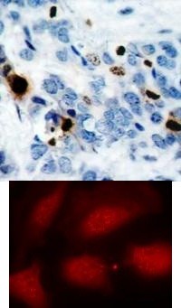
Top Image: Immunohistochemical analysis of paraffin-embedded human breast carcinoma tissue using Histone H3.1 (Phospho-Ser10). Bottom Image: Immunofluorescence staining of methanol-fixed HeLa cells using Histone H3.1 (Phospho-Ser10).Application
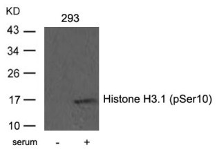
Western blot analysis of lysed extracts from 293 cells untreated or treated with serum using Histone H3.1 (Phospho-Ser10).| Product Name | Histone H3.1 (phospho Ser10) Antibody |
|---|---|
| Antibody Type | Primary Antibodies |
| Product description | Core component of nucleosome. Nucleosomes wrap and compact DNA into chromatin, limiting DNA accessibility to the cellular machineries which require DNA as a template. Histones thereby play a central role in transcription regulation, DNA repair, DNA replication and chromosomal stability. DNA accessibility is regulated via a complex set of post-translational modifications of histones, also called histone code, and nucleosome remodeling.1) Dai J, et al. (2005) Genes Dev 19 (4): 472-488. Yih LH, et al. (2005) Carcinogenesis 26 (1): 53-63. |
| Immunogen | Histone H3.1 (Phospho-Ser10) antibody was raised against a peptide sequence around phosphorylation site of serine 10 (R-K-S (p) -T-G) derived from Human Histone H3.1. |
| Clonality | Polyclonal |
|---|---|
| Host Species | Rabbit |
| Tested Applications | IFIHCWB |
| Western Blot: 1:500~1:1000, Immunohistochemistry: 1:50~1:100, Immunofluorescence: 1:100~1:200 | |
| Species Reactivity | HumanMouseRat |
| Concentration | 1mg/ml |
| Purification | Affinity purified |
| Gene Symbol | HIST1H3D |
|---|---|
| Alternative Names | H3/b H3FB ERBB ERBB1 HER1 H3/a H3/c H3/d H3/f H3/h |
| Molecular Weight(MW) | 17 kDa |
| Tissue Specificity | This antibody detects endogenous level of Histone H3.1 onlywhen phosphorylated at serine 10. |
| Entrez Gene | 8351 |
|---|---|
| Protein Accession | NP_003521.2 |
Application
Top Image: Immunohistochemical analysis of paraffin-embedded human breast carcinoma tissue using Histone H3.1 (Phospho-Ser10). Bottom Image: Immunofluorescence staining of methanol-fixed HeLa cells using Histone H3.1 (Phospho-Ser10).
Application
Western blot analysis of lysed extracts from 293 cells untreated or treated with serum using Histone H3.1 (Phospho-Ser10).| Application Notes | Western Blot: 1:500~1:1000, Immunohistochemistry: 1:50~1:100, Immunofluorescence: 1:100~1:200 |
|---|
| Form | Liquid |
|---|---|
| Storage Instructions | Store antibody at -20˚C for up to one year. |
| Storage Buffer | Antibody supplied in phosphate buffered saline (without Mg2+ and Ca2+), pH 7.4, 150mM NaCl, 0.02% sodium azide and 50% glycerol. |


 Data sheet for OM280987