Application
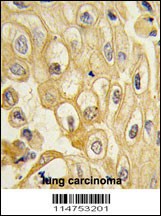
Formalin-fixed and paraffin-embedded human lung carcinoma tissue reacted with the ROR1 antibody , which was peroxidase-conjugated to the secondary antibody, followed by DAB staining.Application
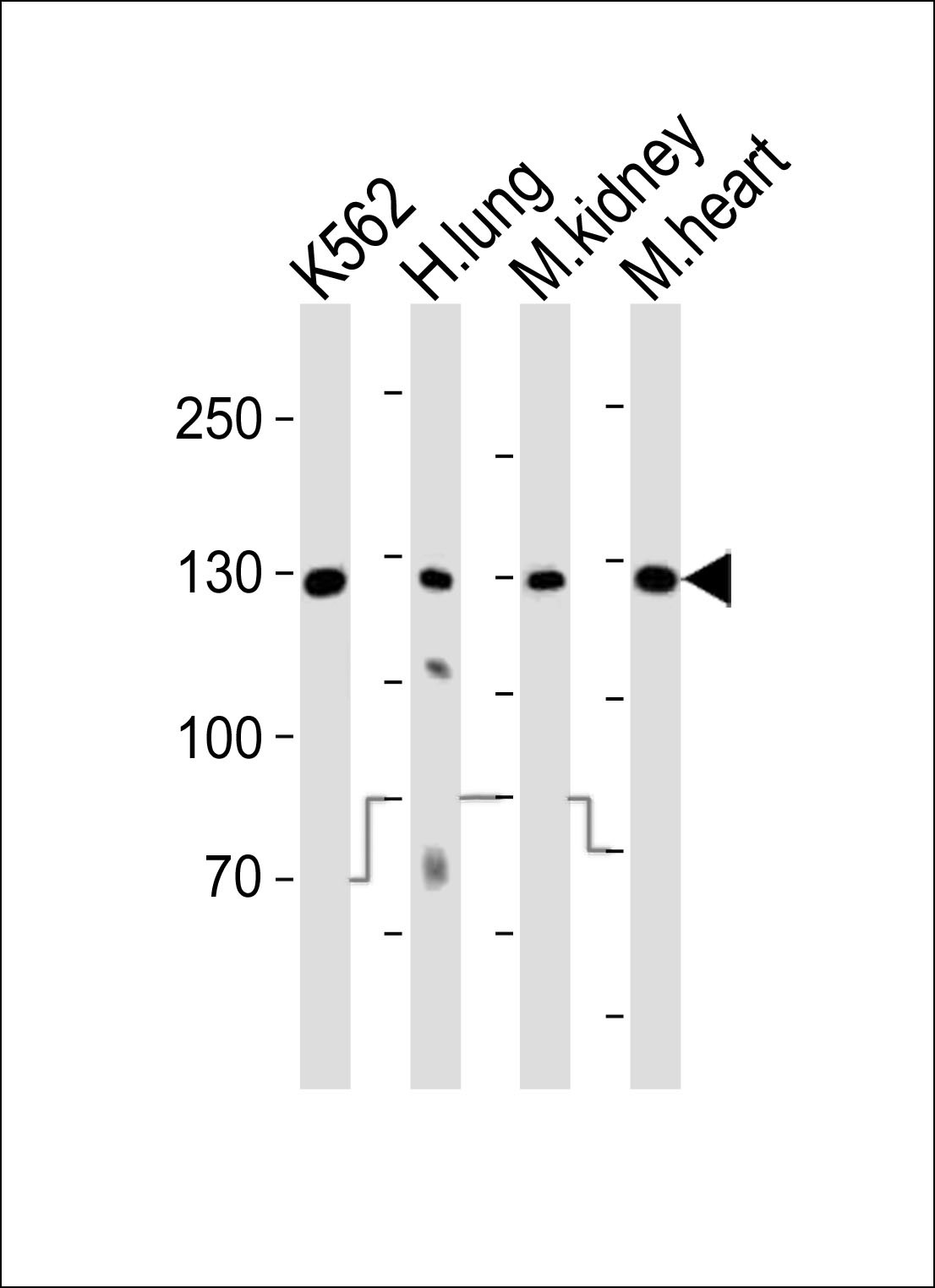
Western blot analysis of lysates from K562 cell line, human lung, mouse kidney, mouse heart tissue (from left to right), using ROR1 Antibody at 1:1000 at each lane.Application
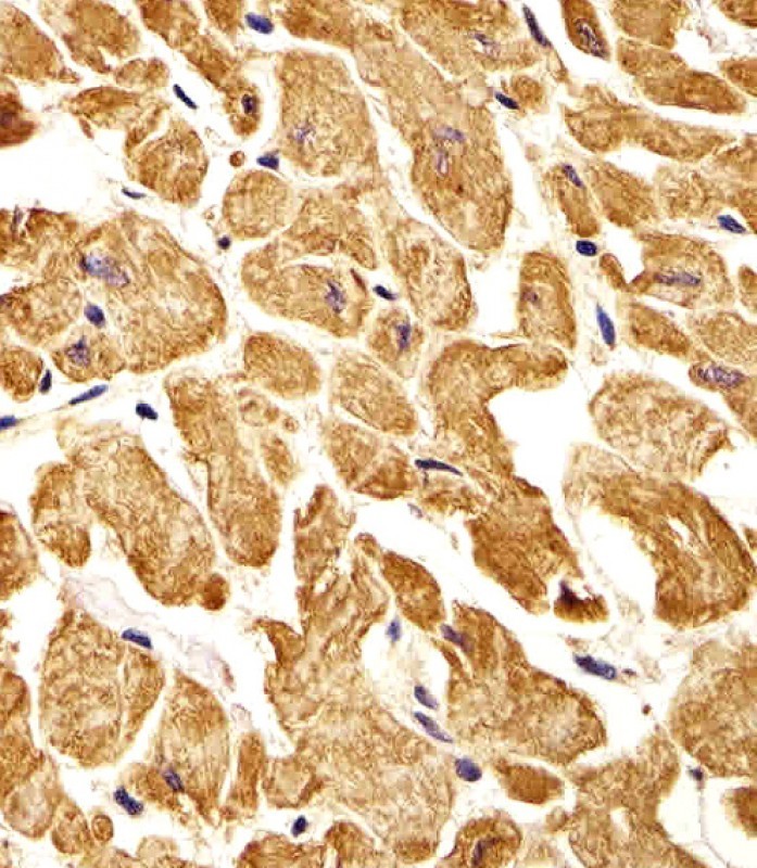
Immunohistochemical analysis of paraffin-embedded H. heart section using ROR1 Antibody . Antibody was diluted at 1:25 dilution. A undiluted biotinylated goat polyvalent antibody was used as the secondary, followed by DAB staining.Application
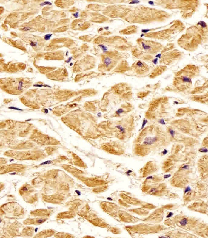
Antibody staining ROR1 in Human heart tissue sections by Immunohistochemistry (IHC-P - paraformaldehyde-fixed, paraffin-embedded sections).Application
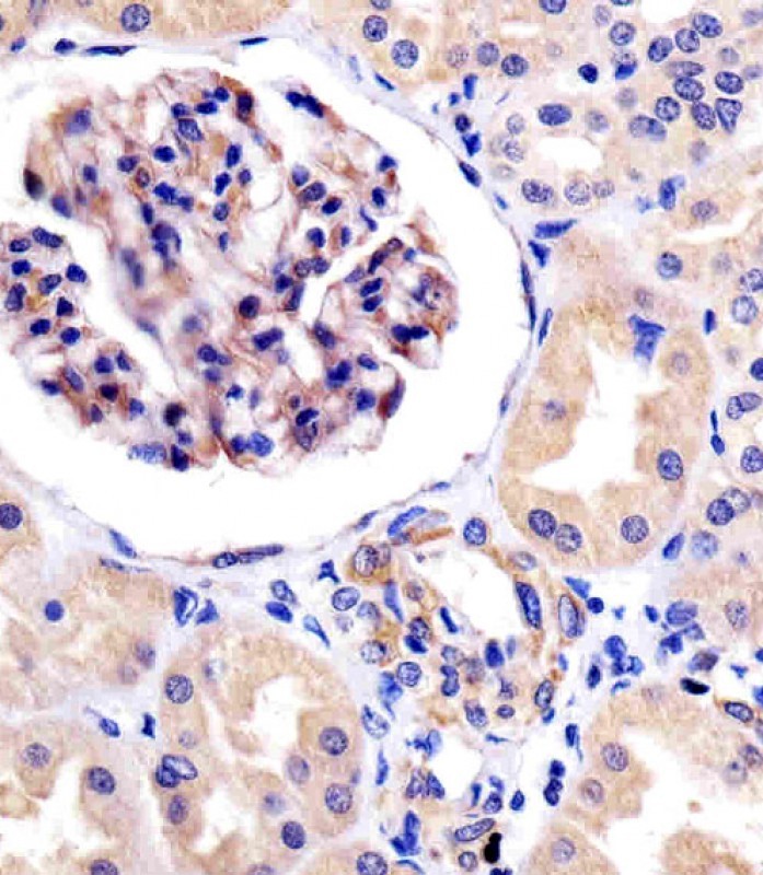
Antibody staining ROR1 in human kidney tissue sections by Immunohistochemistry (IHC-P - paraformaldehyde-fixed, paraffin-embedded sections).Application
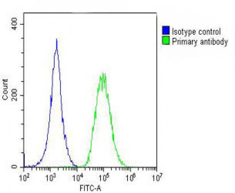
Overlay histogram showing A549 cells stained with Antibody (green line). The cells were fixed with 2% paraformaldehyde (10 min). The cells were then icubated in 2% bovine serum albumin to block non-specific protein-protein interactions followed by the antibody (1:25 dilution) for 60 min at 37ºC. The secondary antibody used was Goat-Anti-Rabbit IgG, DyLight 488 Conjugated Highly Cross-Adsorbed(OH19| Product Name | ROR1 Antibody |
|---|---|
| Antibody Type | Primary Antibodies |
| Antigen Alias | Tyrosine-protein kinase transmembrane receptor ROR1, Neurotrophic tyrosine kinase, receptor-related 1, ROR1, NTRKR1 |
| Product description | ROR1 is a receptor protein tyrosine kinase whose cellular role has not been determined. It is a type I membrane protein and belongs to the ROR subfamily of cell surface receptors. Studies of a similar protein in mouse suggest that this protein may interact with another receptor protein tyrosine kinase and may be involved in skeletal and cardiac development.1) Nomi, M., et al., Mol. Cell. Biol. 21(24):8329-8335 (2001). |
| Immunogen | This ROR1 antibody is generated from rabbits immunized with recombinant human ROR1 protein (aa region: 112 - 399). |
| Clonality | Polyclonal |
|---|---|
| Isotype | Ig |
| Host Species | Rabbit |
| Tested Applications | FACSIHC-PWB |
| For FACS starting dilution is: 1:25 For IHC-P starting dilution is: 1:25 For WB starting dilution is: 1:1000: |
|
| Species Reactivity | HumanMouse |
| Concentration | 1mg/ml |
| Purification | Affinity purified |
| Gene Symbol | ROR1 |
|---|---|
| Alternative Names | Tyrosine-protein kinase transmembrane receptor ROR1 Neurotrophic tyrosine kinase receptor-related 1 ROR1 NTRKR1 |
| Molecular Weight(MW) | 104 kDa |
Application
Formalin-fixed and paraffin-embedded human lung carcinoma tissue reacted with the ROR1 antibody , which was peroxidase-conjugated to the secondary antibody, followed by DAB staining.
Application
Western blot analysis of lysates from K562 cell line, human lung, mouse kidney, mouse heart tissue (from left to right), using ROR1 Antibody at 1:1000 at each lane.
Application
Immunohistochemical analysis of paraffin-embedded H. heart section using ROR1 Antibody . Antibody was diluted at 1:25 dilution. A undiluted biotinylated goat polyvalent antibody was used as the secondary, followed by DAB staining.
Application
Antibody staining ROR1 in Human heart tissue sections by Immunohistochemistry (IHC-P - paraformaldehyde-fixed, paraffin-embedded sections).
Application
Antibody staining ROR1 in human kidney tissue sections by Immunohistochemistry (IHC-P - paraformaldehyde-fixed, paraffin-embedded sections).
Application
Overlay histogram showing A549 cells stained with Antibody (green line). The cells were fixed with 2% paraformaldehyde (10 min). The cells were then icubated in 2% bovine serum albumin to block non-specific protein-protein interactions followed by the antibody (1:25 dilution) for 60 min at 37ºC. The secondary antibody used was Goat-Anti-Rabbit IgG, DyLight 488 Conjugated Highly Cross-Adsorbed(OH19| Application Notes | For FACS starting dilution is: 1:25 For IHC-P starting dilution is: 1:25 For WB starting dilution is: 1:1000: |
|---|
| Form | Liquid |
|---|---|
| Storage Instructions | Store at 4˚C for three months and -20˚C, stable for up to one year. As with all antibodies care should be taken to avoid repeated freeze thaw cycles. Antibodies should not be exposed to prolonged high temperatures. |
| Storage Buffer | Supplied in PBS with 0.09% (W/V) sodium azide. |


 Data sheet for OM290231