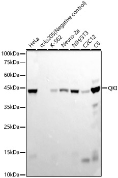

WB
Western blot analysis of various lysates, using QKI Rabbit mAb at 1:20000 dilution. Secondary antibody: HRP Goat Anti-Rabbit IgG (H+L) at 1:10000 dilution. Lysates/proteins: 25ug per lane. Blocking buffer: 3% nonfat dry milk in TBST. Detection: ECL Basic Kit. Exposure time: 30s.IHC
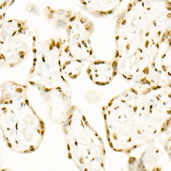
Immunohistochemistry analysis of paraffin embedded Human placenta using QKI Rabbit mAb at dilution of 1:200 (40x lens). High pressure antigen retrieval performed with 0.01M Citrate Bufferr (pH 6.0) prior to IHC staining.ICC/IF
Confocal imaging of NIH-3T3 cells using QKI Rabbit mAb (dilution 1:200)(Red). The cells were counterstained with α-Tubulin Mouse mAb (dilution 1:400)(Green). DAPI was used for nuclear staining (blue). objective: 60x.| Product Name | QKI Rabbit mAb |
|---|---|
| Antibody Type | Primary Antibodies |
| Immunogen | Recombinant fusion protein containing a sequence corresponding to amino acids 1-341 of human QKI. (NP_006766.1). |
| Clonality | Monoclonal |
|---|---|
| Isotype | IgG |
| Host Species | Rabbit |
| Tested Applications | ICC/IFIHCWB |
| WB:1:2000-1:20000 IHC:1:50-1:200 ICC:1:50-1:200 |
|
| Species Reactivity | HumanMouseRat |
| Concentration | 1mg/ml |
| Purification | Affinity purified |
| Gene Symbol | QKI |
|---|---|
| Gene Synonyms | QK Hqk QK1 QK3 hqkI |
| Gene Full Name | QKI, KH domain containing RNA binding |
| Gene Summary | The protein encoded by this gene is an RNA-binding protein that regulates pre-mRNA splicing, export of mRNAs from the nucleus, protein translation, and mRNA stability. The encoded protein is involved in myelinization and oligodendrocyte differentiation and may play a role in schizophrenia. Multiple transcript variants encoding different isoforms have been found for this gene. [provided by RefSeq, Jul 2014] |
| Molecular Weight(MW) | 38kDa(Observed MW 42kDa) |
| Cellular Localization | Cytoplasm,Nucleus. |
WB
Western blot analysis of various lysates, using QKI Rabbit mAb at 1:20000 dilution. Secondary antibody: HRP Goat Anti-Rabbit IgG (H+L) at 1:10000 dilution. Lysates/proteins: 25ug per lane. Blocking buffer: 3% nonfat dry milk in TBST. Detection: ECL Basic Kit. Exposure time: 30s.
IHC
Immunohistochemistry analysis of paraffin embedded Human placenta using QKI Rabbit mAb at dilution of 1:200 (40x lens). High pressure antigen retrieval performed with 0.01M Citrate Bufferr (pH 6.0) prior to IHC staining.
ICC/IF
Confocal imaging of NIH-3T3 cells using QKI Rabbit mAb (dilution 1:200)(Red). The cells were counterstained with α-Tubulin Mouse mAb (dilution 1:400)(Green). DAPI was used for nuclear staining (blue). objective: 60x.| Application Notes | WB:1:2000-1:20000 IHC:1:50-1:200 ICC:1:50-1:200 |
|---|
| Form | Liquid |
|---|---|
| Storage Instructions | Store at -20℃. Avoid freeze / thaw cycles. |
| Storage Buffer | Buffer: PBS with 0.05% proclin300,0.05% BSA,50% glycerol,pH7.3. |


 Data sheet for OM643374