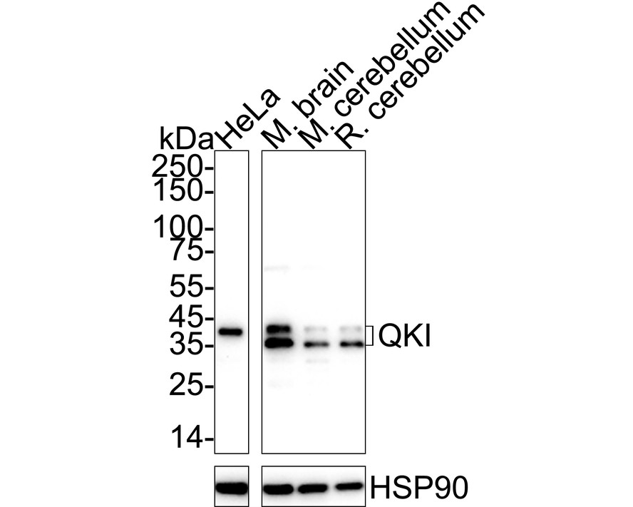
WB
Western blot analysis of QKI on different lysates with Mouse anti-QKI antibody at 1/2,000 dilution. Lane 1: HeLa cell lysate, Lane 2: Mouse brain tissue lysate, Lane 3: Mouse cerebellum tissue lysate, Lane 4: Rat cerebellum tissue lysate, Lysates/proteins at 30 µg/Lane. Exposure time: 3 minutes; 4-20% SDS-PAGE gel. Proteins were transferred to a PVDF membrane and blocked with 5% NFDM/TBST for 1 hour at room temperature. The primary antibody at 1/2,000 dilution was used in 5% NFDM/TBST at 4℃ overnight. Goat Anti-Mouse IgG - HRP Secondary Antibody at 1/50,000 dilution was used for 1 hour at room temperature.IHC
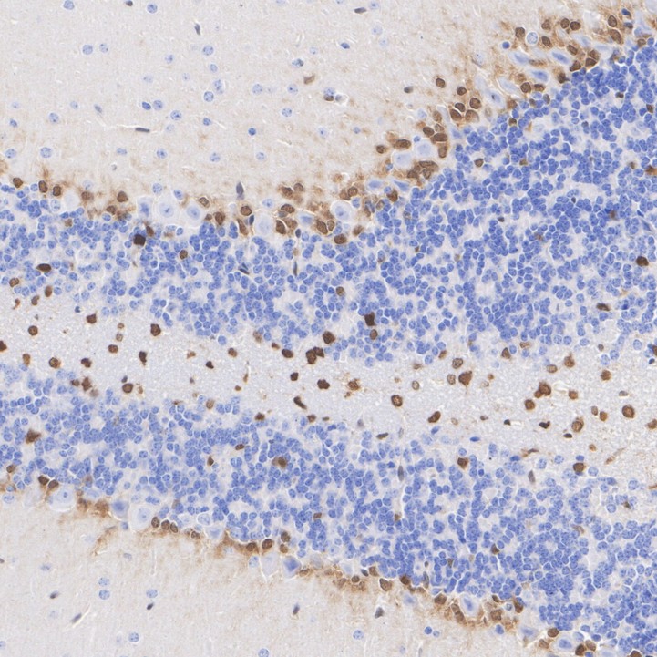
Immunohistochemical analysis of paraffin-embedded mouse cerebellum tissue with Mouse anti-QKI antibody at 1/200 dilution. The section was pre-treated using heat mediated antigen retrieval with Tris-EDTA buffer (pH 9.0) for 20 minutes. The tissues were blocked in 1% BSA for 20 minutes at room temperature, washed with ddH2O and PBS, and then probed with the primary antibody at 1/200 dilution for 1 hour at room temperature. The detection was performed using an HRP conjugated compact polymer system. DAB was used as the chromogen. Tissues were counterstained with hematoxylin and mounted with DPX.ICC/IF
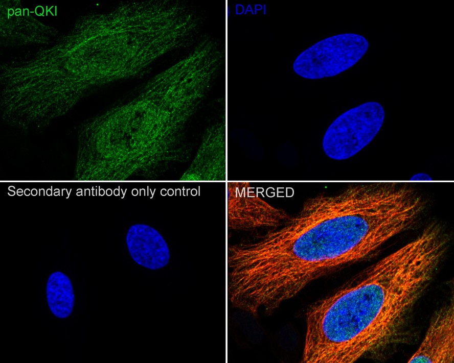
Immunocytochemistry analysis of HeLa cells labeling QKI with Mouse anti-QKI antibody at 1/100 dilution. Cells were fixed in 4% paraformaldehyde for 15 minutes at room temperature, permeabilized with 0.1% Triton X-100 in PBS for 15 minutes at room temperature, then blocked with 1% BSA in 10% negative goat serum for 1 hour at room temperature. Cells were then incubated with Mouse anti-QKI antibody at 1/100 dilution in 1% BSA in PBST overnight at 4 ℃. Goat Anti-Mouse IgG H&L (iFluor™ 488) was used as the secondary antibody at 1/1,000 dilution. PBS instead of the primary antibody was used as the secondary antibody only control. Nuclear DNA was labelled in blue with DAPI. beta Tubulin (red) was stained at 1/100 dilution overnight at +4℃. Goat Anti-Rabbit IgG H&L (iFluor™ 594) were used as the secondary antibody at 1/1,000 dilution.IF-F
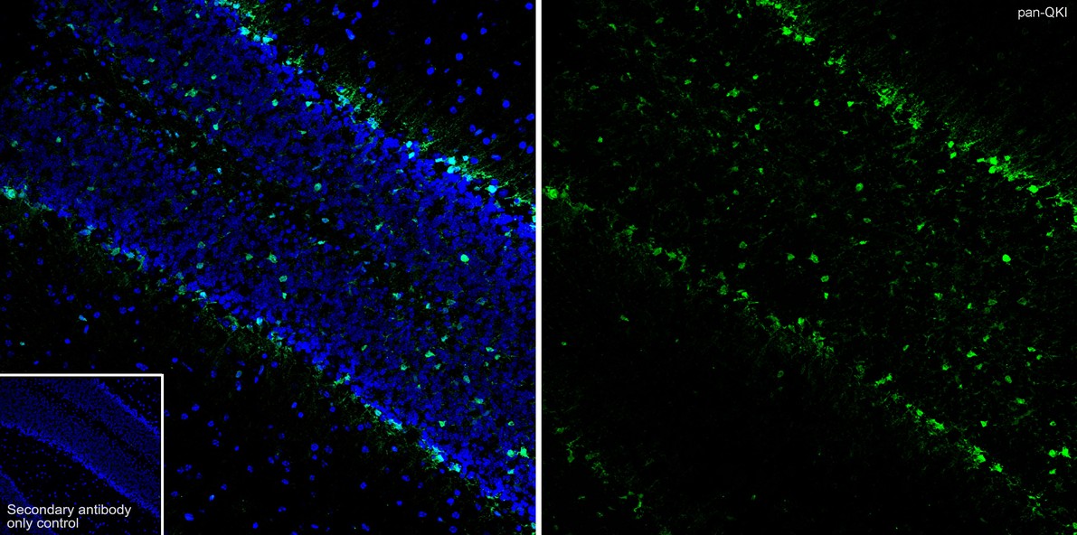
Immunofluorescence analysis of frozen mouse cerebellum tissue with Mouse anti-QKI antibody at 1/500 dilution. The section was not undergone antigen retrieval. The tissues were blocked in 10% negative goat serum for 1 hour at room temperature, washed with PBS, and then probed with the primary antibody (green) at 1/500 dilution overnight at 4 ℃, washed with PBS. Goat Anti-Mouse IgG H&L (iFluor™ 488) was used as the secondary antibody at 1/1,000 dilution. Nuclei were counterstained with DAPI (blue).IP
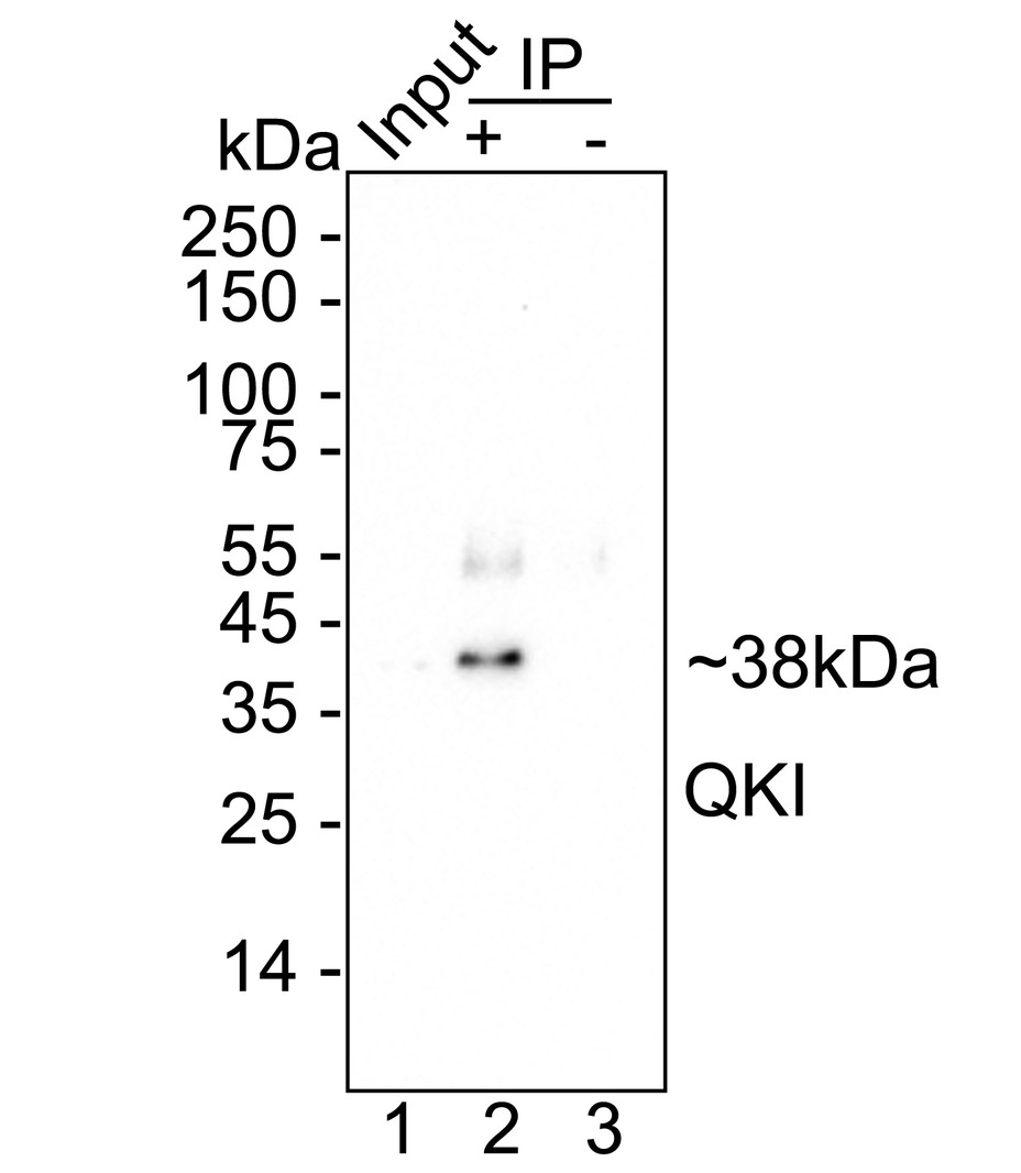
QKI was immunoprecipitated from 0.2 mg HeLa cell lysate with Mouse anti-QKI antibody at 2 µg/10 µl beads. Western blot was performed from the immunoprecipitate using Mouse anti-QKI antibody at 1/1,000 dilution. Anti-Mouse IgG for IP Nano-secondary antibody at 1/5,000 dilution was used for 1 hour at room temperature. Lane 1: HeLa cell lysate (input), Lane 2: Mouse anti-QKI antibody IP in HeLa cell lysate, Lane 3: Mouse IgG instead of Mouse anti-QKI antibody in HeLa cell lysate. Blocking/Dilution buffer: 5% NFDM/TBST. Exposure time: 3 minutes.| Product Name | QKI Recombinant Mouse Monoclonal Antibody |
|---|---|
| Antibody Type | Primary Antibodies |
| Immunogen | Recombinant protein within human QKI aa 1-341 / 341. |
| Clonality | monoclonal |
|---|---|
| Isotype | IgG1 |
| Host Species | Mouse |
| Tested Applications | ICC/IFIF-FIHCIPWB |
| WB:1:2000 IHC:1:200 ICC/IF:1:100 IF-F:1:500 IP:1-2μg/sample |
|
| Species Reactivity | HumanMouseRat |
| Concentration | 1mg/ml |
| Purification | Protein A |
| Gene Symbol | QKI |
|---|---|
| Gene Synonyms | QK Hqk QK1 QK3 hqkI |
| Gene Full Name | QKI, KH domain containing RNA binding |
| Gene Summary | The protein encoded by this gene is an RNA-binding protein that regulates pre-mRNA splicing, export of mRNAs from the nucleus, protein translation, and mRNA stability. The encoded protein is involved in myelinization and oligodendrocyte differentiation and may play a role in schizophrenia. Multiple transcript variants encoding different isoforms have been found for this gene. [provided by RefSeq, Jul 2014] |
| Molecular Weight(MW) | 38kDa(Observed band size: 35-38kDa) |
| Cellular Localization | Nucleus, Cytoplasm. |
WB
Western blot analysis of QKI on different lysates with Mouse anti-QKI antibody at 1/2,000 dilution. Lane 1: HeLa cell lysate, Lane 2: Mouse brain tissue lysate, Lane 3: Mouse cerebellum tissue lysate, Lane 4: Rat cerebellum tissue lysate, Lysates/proteins at 30 µg/Lane. Exposure time: 3 minutes; 4-20% SDS-PAGE gel. Proteins were transferred to a PVDF membrane and blocked with 5% NFDM/TBST for 1 hour at room temperature. The primary antibody at 1/2,000 dilution was used in 5% NFDM/TBST at 4℃ overnight. Goat Anti-Mouse IgG - HRP Secondary Antibody at 1/50,000 dilution was used for 1 hour at room temperature.
IHC
Immunohistochemical analysis of paraffin-embedded mouse cerebellum tissue with Mouse anti-QKI antibody at 1/200 dilution. The section was pre-treated using heat mediated antigen retrieval with Tris-EDTA buffer (pH 9.0) for 20 minutes. The tissues were blocked in 1% BSA for 20 minutes at room temperature, washed with ddH2O and PBS, and then probed with the primary antibody at 1/200 dilution for 1 hour at room temperature. The detection was performed using an HRP conjugated compact polymer system. DAB was used as the chromogen. Tissues were counterstained with hematoxylin and mounted with DPX.
ICC/IF
Immunocytochemistry analysis of HeLa cells labeling QKI with Mouse anti-QKI antibody at 1/100 dilution. Cells were fixed in 4% paraformaldehyde for 15 minutes at room temperature, permeabilized with 0.1% Triton X-100 in PBS for 15 minutes at room temperature, then blocked with 1% BSA in 10% negative goat serum for 1 hour at room temperature. Cells were then incubated with Mouse anti-QKI antibody at 1/100 dilution in 1% BSA in PBST overnight at 4 ℃. Goat Anti-Mouse IgG H&L (iFluor™ 488) was used as the secondary antibody at 1/1,000 dilution. PBS instead of the primary antibody was used as the secondary antibody only control. Nuclear DNA was labelled in blue with DAPI. beta Tubulin (red) was stained at 1/100 dilution overnight at +4℃. Goat Anti-Rabbit IgG H&L (iFluor™ 594) were used as the secondary antibody at 1/1,000 dilution.
IF-F
Immunofluorescence analysis of frozen mouse cerebellum tissue with Mouse anti-QKI antibody at 1/500 dilution. The section was not undergone antigen retrieval. The tissues were blocked in 10% negative goat serum for 1 hour at room temperature, washed with PBS, and then probed with the primary antibody (green) at 1/500 dilution overnight at 4 ℃, washed with PBS. Goat Anti-Mouse IgG H&L (iFluor™ 488) was used as the secondary antibody at 1/1,000 dilution. Nuclei were counterstained with DAPI (blue).
IP
QKI was immunoprecipitated from 0.2 mg HeLa cell lysate with Mouse anti-QKI antibody at 2 µg/10 µl beads. Western blot was performed from the immunoprecipitate using Mouse anti-QKI antibody at 1/1,000 dilution. Anti-Mouse IgG for IP Nano-secondary antibody at 1/5,000 dilution was used for 1 hour at room temperature. Lane 1: HeLa cell lysate (input), Lane 2: Mouse anti-QKI antibody IP in HeLa cell lysate, Lane 3: Mouse IgG instead of Mouse anti-QKI antibody in HeLa cell lysate. Blocking/Dilution buffer: 5% NFDM/TBST. Exposure time: 3 minutes.| Application Notes | WB:1:2000 IHC:1:200 ICC/IF:1:100 IF-F:1:500 IP:1-2μg/sample |
|---|
| Form | Liquid |
|---|---|
| Storage Instructions | Store at +4℃ after thawing. Aliquot store at -20℃ or -80℃. Avoid repeated freeze / thaw cycles. |
| Storage Buffer | 1*TBS (pH7.4), 0.05% BSA, 40% Glycerol. Preservative: 0.05% Sodium Azide. |


 Data sheet for OM643928