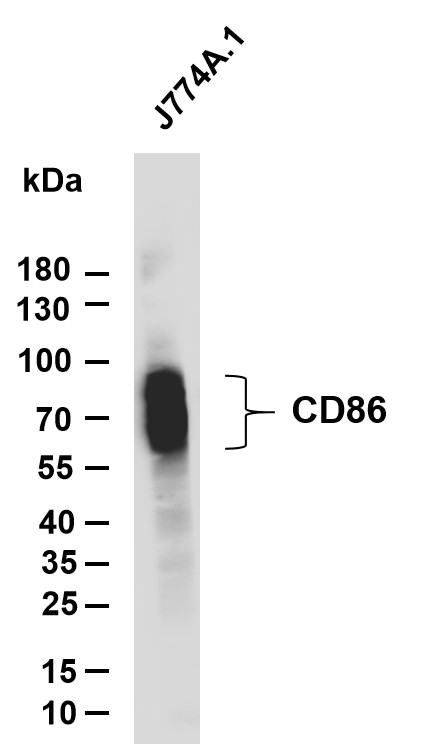

WB
Various whole cell lysates were separated by 4-20% SDS-PAGE, and the membrane was blotted with anti-CD86 antibody. The HRP- conjugated Goat anti-Rabbit IgG(H + L) antibody was used to detect the antibody. Lane 1: J774A.1 Predicted band size: 35kDa Observed band size:60-85kDaIHC
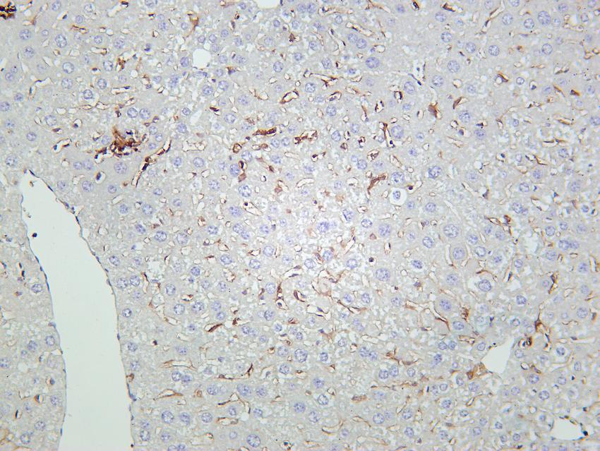
Mouse liver tissue was stained with Anti-CD86 rabbit AntibodyIHC
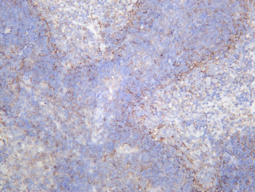
Mouse spleen was stained with anti-CD86 rabbit antibodyIF
Immunofluorescence analysis of paraffin-embedded Mouse spleen. Primary Antibody was diluted at 1:200(4° overnight). an Multi colour-Fluorescence kit . EDTA based antigen retrieval was used before Green tyramide signal amplification. DAPI (dark blue) was used as a nuclear counter stain. Microscopy and pseudocoloring of individual dyes was performed using a Slideviewer Imaging System (3D histech).| Product Name | CD86 Rabbit mAb |
|---|---|
| Antibody Type | Primary Antibodies |
| Product description | This gene encodes a type I membrane protein that is a member of the immunoglobulin superfamily. This protein is expressed by antigen-presenting cells, and it is the ligand for two proteins at the cell surface of T cells, CD28 antigen and cytotoxic T-lymphocyte-associated protein 4. Binding of this protein with CD28 antigen is a costimulat Function:Receptor involved in the costimulatory signal essential for T-lymphocyte proliferation and interleukin-2 production, by binding CD28 or CTLA-4. May play a critical role in the early events of T-cell activation and costimulation of naive T-cells, such as deciding between immunity and anergy that is made by T-cells within 24 hours after activation. Isoform 2 interferes with the formation of CD86 clusters, and thus acts as a negative regulator of T-cell activation.,online information:CD86 entry,PTM:Polyubiquitinated; which is promoted by MARCH8 and results in endocytosis and lysosomal degradation.,similarity:Contains 1 Ig-like C2-type (immunoglobulin-like) domain.,similarity:Contains 1 Ig-like V-type (immunoglobulin-like) domain.,subunit:Interacts with MARCH8. Interacts with human herpesvirus 8 MIR2 protein (Probable). Interacts with adenovirus subgroup B fiber proteins and acts as a receptor for these viruses.,tissue specificity:Expressed by activated B-lymphocytes and monocytes., |
| Clonality | Monoclonal |
|---|---|
| Isotype | IgG,K |
| Host Species | Rabbit |
| Tested Applications | ELISAICC/IFIHCWB |
| WB:1:500-1:2000 IHC:1:200-1:1000 ICC:1:100-1:500 IP:1:50-1:200 ELISA 1:5000-20000: |
|
| Species Reactivity | Mouse |
| Concentration | 1mg/ml |
| Purification | Protein A |
| Gene Symbol | CD86 |
|---|---|
| Gene Synonyms | B7 B70 MB7 B7-2 B7.2 CLS1 Ly58 ETC-1 Ly-58 MB7-2 Cd28l2 TS/A-2 |
| Gene Full Name | Cd86 CD86 antigen [ Mus musculus (house mouse) ] |
| Gene Summary | Predicted to enable receptor ligand activity. Involved in several processes, including positive regulation of immunoglobulin production; positive regulation of non-canonical NF-kappaB signal transduction; and toll-like receptor 3 signaling pathway. Acts upstream of or within several processes, including cellular response to lipopolysaccharide; positive regulation of T cell proliferation; and toll-like receptor signaling pathway. Located in external side of plasma membrane and intracellular membrane-bounded organelle. Is expressed in central nervous system; mandible; and retina. Used to study Guillain-Barre syndrome. Human ortholog(s) of this gene implicated in several diseases, including Henoch-Schoenlein purpura; autoimmune disease (multiple); chronic lymphocytic leukemia; chronic obstructive pulmonary disease; and systemic scleroderma. Orthologous to human CD86 (CD86 molecule). [provided by Alliance of Genome Resources, Dec 2024] |
| Alternative Names | Activation B7-2 antigen B70 BU63 CTLA-4 counter-receptor B7.2 FUN-1 |
| Molecular Weight(MW) | 60-80kD (Calculated) |
| Cellular Localization | Membranous |
WB
Various whole cell lysates were separated by 4-20% SDS-PAGE, and the membrane was blotted with anti-CD86 antibody. The HRP- conjugated Goat anti-Rabbit IgG(H + L) antibody was used to detect the antibody. Lane 1: J774A.1 Predicted band size: 35kDa Observed band size:60-85kDa
IHC
Mouse liver tissue was stained with Anti-CD86 rabbit Antibody
IHC
Mouse spleen was stained with anti-CD86 rabbit antibody
IF
Immunofluorescence analysis of paraffin-embedded Mouse spleen. Primary Antibody was diluted at 1:200(4° overnight). an Multi colour-Fluorescence kit . EDTA based antigen retrieval was used before Green tyramide signal amplification. DAPI (dark blue) was used as a nuclear counter stain. Microscopy and pseudocoloring of individual dyes was performed using a Slideviewer Imaging System (3D histech).| Application Notes | WB:1:500-1:2000 IHC:1:200-1:1000 ICC:1:100-1:500 IP:1:50-1:200 ELISA 1:5000-20000: |
|---|
| Form | Liquid |
|---|---|
| Storage Instructions | Shipped at 4°C. Store at +4°C short term (1-2 weeks). Store at -20°C long term. Avoid freeze / thaw cycle. |
| Storage Buffer | Purified antibody in PBS with 0.05% sodium azide |


 Data sheet for OM643454