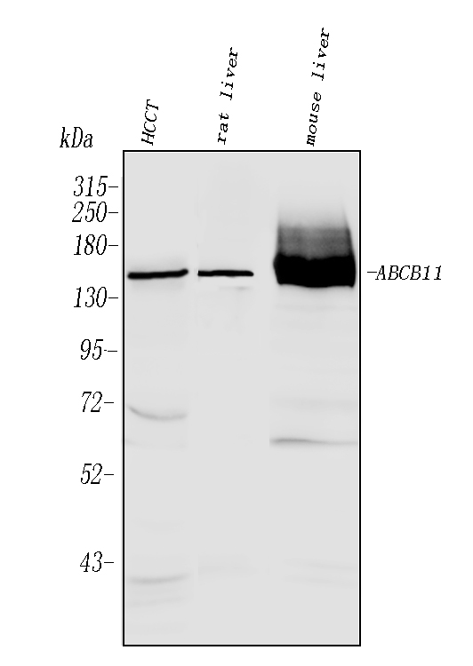
WB
Western blot analysis of BSEP/ABCB11 using anti-BSEP/ABCB11 antibody. The sample well of each lane was loaded with 30 ug of sample under reducing conditions. Lane 1: HCCT tissue lysates, Lane 2: rat liver tissue lysates, Lane 3: mouse liver tissue lysates. After electrophoresis, proteins were transferred to a membrane. Then the membrane was incubated with rabbit anti-BSEP/ABCB11 antigen affinity purified polyclonal antibody at a dilution of 1:1000 and probed with a goat anti-rabbit IgG-HRP secondary antibody. The signal is developed using ECL Plus Western Blotting Substrate.IHC
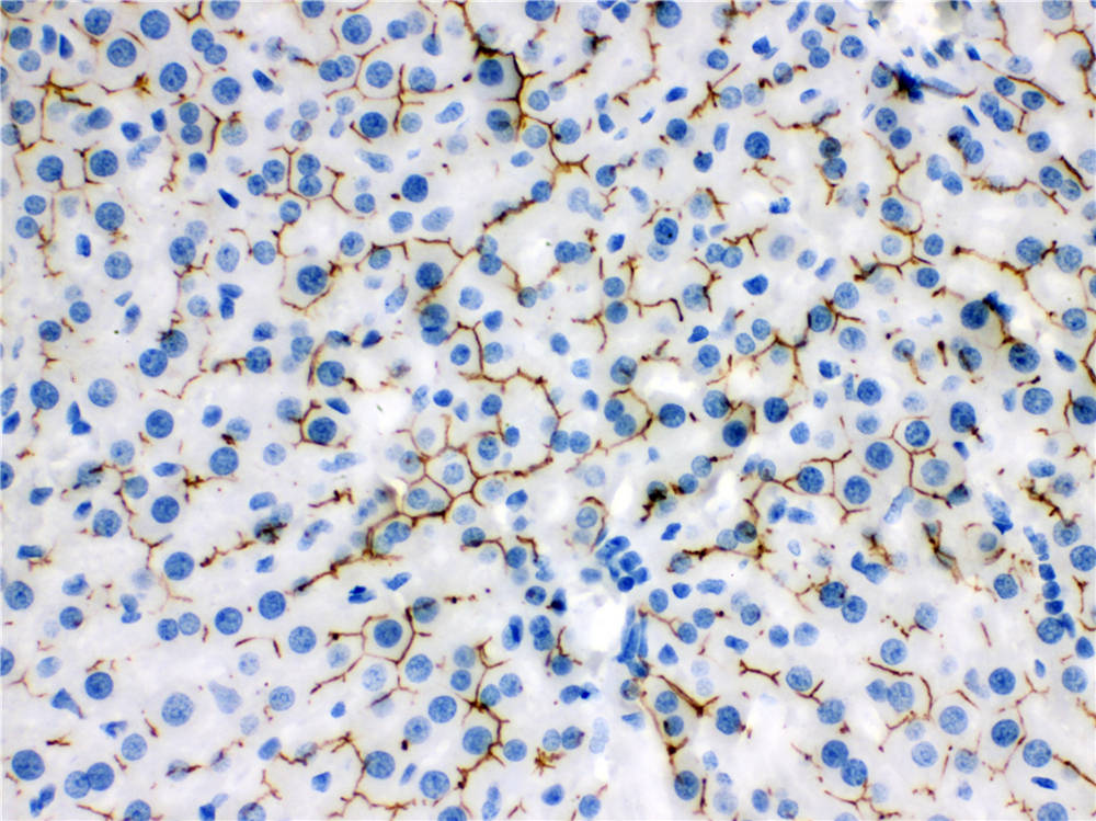
IHC analysis of BSEP/ABCB11 using anti-BSEP/ABCB11 antibody. BSEP/ABCB11 was detected in a paraffin-embedded section of rat liver tissue. Biotinylated goat anti-rabbit IgG was used as secondary antibody. The tissue section was incubated with rabbit anti-BSEP/ABCB11 Antibody at a dilution of 1:200 and developed using Strepavidin-Biotin-Complex (SABC) with DAB as the chromogen.IF-P
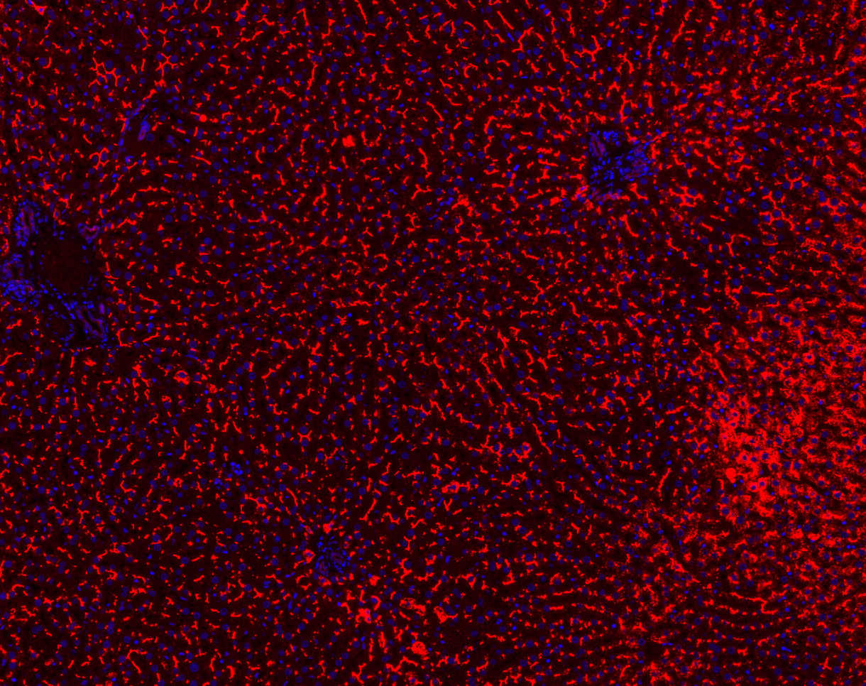
IF analysis of ABCB11 using anti- ABCB11 antibody ABCB11 was detected in paraffin-embedded section of mouse liver tissues. Heat mediated antigen retrieval was performed in citrate buffer (pH6, epitope retrieval solution ) for 20 mins. The tissue section was blocked with 10% goat serum. The tissue section was then incubated with 1μg/ml rabbit anti- ABCB11 Antibody overnight at 4°C. Cy3 Conjugated Goat Anti-Rabbit IgG was used as secondary antibody at 1:100 dilution and incubated for 30 minutes at 37°C. The section was counterstained with DAPI. Visualize using a fluorescence microscope and filter sets appropriate for the label used.| Product Name | Rabbit polyclonal antibody to BSEP/ABCB11 |
|---|---|
| Antibody Type | Primary Antibodies |
| Immunogen | A synthetic peptide corresponding to a sequence at the C-terminus of human ABCB11, different from the related mouse and rat sequences by three amino acids. |
| Clonality | Polyclonal |
|---|---|
| Isotype | IgG |
| Host Species | Rabbit |
| Tested Applications | IF-PIHCWB |
| WB:1:500-1:2000 IHC:1:50-1:400 IF-P:1:50-1:400 |
|
| Species Reactivity | HumanMouseRat |
| Concentration | 0.5mg/ml |
| Purification | Affinity purified |
| Gene Symbol | ABCB11 |
|---|---|
| Gene Synonyms | BSEP PGY4 SPGP ABC16 BRIC2 PFIC2 PFIC-2 |
| Gene Full Name | ATP binding cassette subfamily B member 11 |
| Gene Summary | The membrane-associated protein encoded by this gene is a member of the superfamily of ATP-binding cassette (ABC) transporters. ABC proteins transport various molecules across extra- and intra-cellular membranes. ABC genes are divided into seven distinct subfamilies (ABC1, MDR/TAP, MRP, ALD, OABP, GCN20, White). This protein is a member of the MDR/TAP subfamily. Members of the MDR/TAP subfamily are involved in multidrug resistance. The protein encoded by this gene is the major canalicular bile salt export pump in man. Mutations in this gene cause a form of progressive familial intrahepatic cholestases which are a group of inherited disorders with severe cholestatic liver disease from early infancy. [provided by RefSeq, Jul 2008] |
| Molecular Weight(MW) | 146kDa |
| Cellular Localization | Membrane, Multi-pass membrane protein. |
WB
Western blot analysis of BSEP/ABCB11 using anti-BSEP/ABCB11 antibody. The sample well of each lane was loaded with 30 ug of sample under reducing conditions. Lane 1: HCCT tissue lysates, Lane 2: rat liver tissue lysates, Lane 3: mouse liver tissue lysates. After electrophoresis, proteins were transferred to a membrane. Then the membrane was incubated with rabbit anti-BSEP/ABCB11 antigen affinity purified polyclonal antibody at a dilution of 1:1000 and probed with a goat anti-rabbit IgG-HRP secondary antibody. The signal is developed using ECL Plus Western Blotting Substrate.
IHC
IHC analysis of BSEP/ABCB11 using anti-BSEP/ABCB11 antibody. BSEP/ABCB11 was detected in a paraffin-embedded section of rat liver tissue. Biotinylated goat anti-rabbit IgG was used as secondary antibody. The tissue section was incubated with rabbit anti-BSEP/ABCB11 Antibody at a dilution of 1:200 and developed using Strepavidin-Biotin-Complex (SABC) with DAB as the chromogen.
IF-P
IF analysis of ABCB11 using anti- ABCB11 antibody ABCB11 was detected in paraffin-embedded section of mouse liver tissues. Heat mediated antigen retrieval was performed in citrate buffer (pH6, epitope retrieval solution ) for 20 mins. The tissue section was blocked with 10% goat serum. The tissue section was then incubated with 1μg/ml rabbit anti- ABCB11 Antibody overnight at 4°C. Cy3 Conjugated Goat Anti-Rabbit IgG was used as secondary antibody at 1:100 dilution and incubated for 30 minutes at 37°C. The section was counterstained with DAPI. Visualize using a fluorescence microscope and filter sets appropriate for the label used.| Application Notes | WB:1:500-1:2000 IHC:1:50-1:400 IF-P:1:50-1:400 |
|---|
| Form | Liquid |
|---|---|
| Storage Instructions | 12 months from date of receipt,-20℃ as supplied. 6 months 2 to 8℃ after reconstitution. Avoid repeated freezing and thawing. |
| Storage Buffer | 500 ug/ml antibody with PBS, 0.02% NaN3, 1 mg/ml BSA and 50% glycerol. |


 Data sheet for OM643709