WB
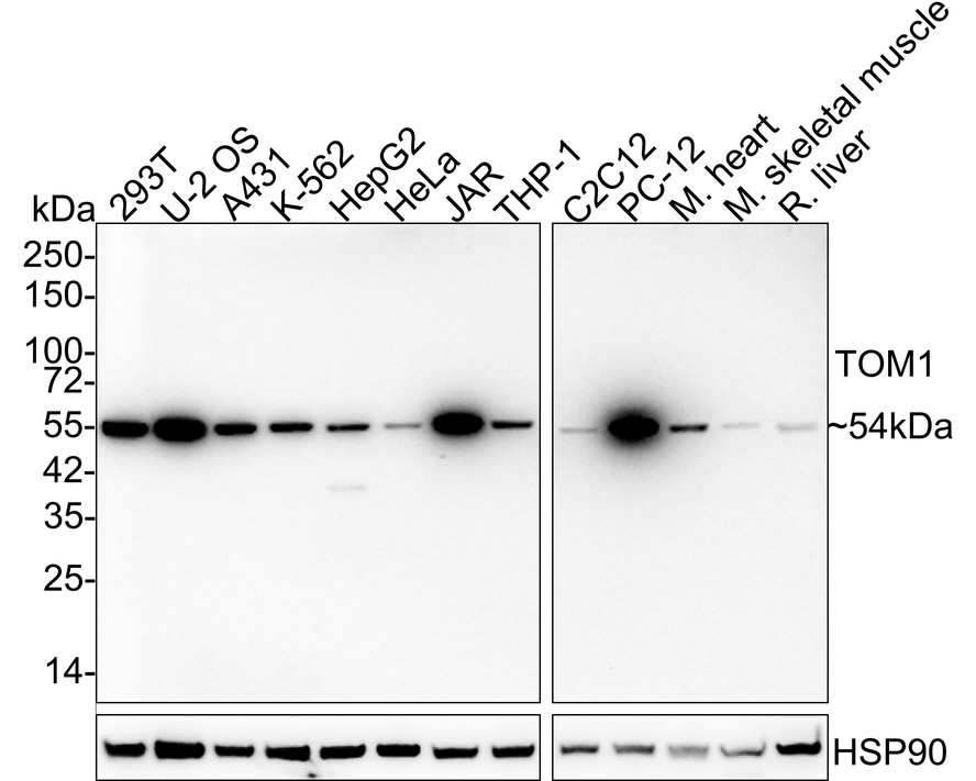
Western blot analysis of TOM1 on different lysates with Rabbit anti-TOM1 antibody at 1/2,000 dilution. Lane 1: 293T cell lysate (20 µg/Lane), Lane 2: U-2 OS cell lysate (20 µg/Lane), Lane 3: A431 cell lysate (20 µg/Lane), Lane 4: K-562 cell lysate (20 µg/Lane), Lane 5: HepG2 cell lysate (20 µg/Lane), Lane 6: HeLa cell lysate (20 µg/Lane), Lane 7: JAR cell lysate (20 µg/Lane), Lane 8: THP-1 cell lysate (20 µg/Lane), Lane 9: C2C12 cell lysate (20 µg/Lane), Lane 10: PC-12 cell lysate (20 µg/Lane), Lane 11: Mouse heart tissue lysate (40 µg/Lane), Lane 12: Mouse skeletal muscle tissue lysate (40 µg/Lane), Lane 13: Rat liver tissue lysate (40 µg/Lane), Exposure time: 3 minutes; 4-20% SDS-PAGE gel. Proteins were transferred to a PVDF membrane and blocked with 5% NFDM/TBST for 1 hour at room temperature. The primary antibody at 1/2,000 dilution was used in 5% NFDM/TBST at 4℃ overnight. Goat Anti-Rabbit IgG - HRP Secondary Antibody at 1/50,000 dilution was used for 1 hour at room temperature.IHC
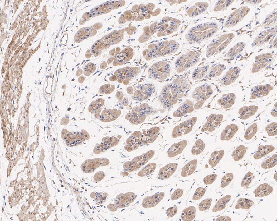
Immunohistochemical analysis of paraffin-embedded mouse stomach tissue with Rabbit anti-TOM1 antibody at 1/200 dilution. The section was pre-treated using heat mediated antigen retrieval with Tris-EDTA buffer (pH 9.0) for 20 minutes. The tissues were blocked in 1% BSA for 20 minutes at room temperature, washed with ddH2O and PBS, and then probed with the primary antibody at 1/200 dilution for 1 hour at room temperature. The detection was performed using an HRP conjugated compact polymer system. DAB was used as the chromogen. Tissues were counterstained with hematoxylin and mounted with DPX.IF-P
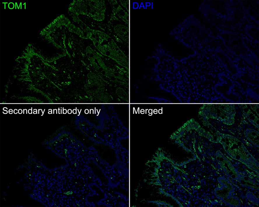
Immunofluorescence analysis of paraffin-embedded human stomach carcinoma tissue labeling TOM1 with Rabbit anti-TOM1 antibody at 1/200 dilution. The section was pre-treated using heat mediated antigen retrieval with Tris-EDTA buffer (pH 9.0) for 20 minutes. The tissues were blocked in 10% negative goat serum for 1 hour at room temperature, washed with PBS, and then probed with the primary antibody (green) at 1/200 dilution overnight at 4 ℃, washed with PBS. Goat Anti-Rabbit IgG H&L (iFluor™ 488) was used as the secondary antibody at 1/1,000 dilution. Nuclei were counterstained with DAPI (blue).| Product Name | TOM1 Recombinant Rabbit Monoclonal Antibody |
|---|---|
| Antibody Type | Primary Antibodies |
| Immunogen | Recombinant protein within human TOM1 aa 300-450. |
| Clonality | monoclonal |
|---|---|
| Isotype | IgG |
| Host Species | Rabbit |
| Tested Applications | IF-PIHCWB |
| WB:1:2000 IHC:1:200-1:1000 IF-P:1:200 |
|
| Species Reactivity | HumanMouseRat |
| Concentration | 1mg/ml |
| Purification | Protein A |
| Gene Symbol | TOM1 |
|---|---|
| Gene Synonyms | IMD85 |
| Gene Full Name | target of myb1 membrane trafficking protein |
| Gene Summary | This gene was identified as a target of the v-myb oncogene. The encoded protein shares its N-terminal domain in common with proteins associated with vesicular trafficking at the endosome. It is recruited to the endosomes by its interaction with endofin. Several alternatively spliced transcript variants encoding different isoforms have been found for this gene. [provided by RefSeq, Oct 2008] |
| Molecular Weight(MW) | 54kDa |
| Cellular Localization | Cytoplasm, Endosome membrane, Early endosome membrane. |
WB
Western blot analysis of TOM1 on different lysates with Rabbit anti-TOM1 antibody at 1/2,000 dilution. Lane 1: 293T cell lysate (20 µg/Lane), Lane 2: U-2 OS cell lysate (20 µg/Lane), Lane 3: A431 cell lysate (20 µg/Lane), Lane 4: K-562 cell lysate (20 µg/Lane), Lane 5: HepG2 cell lysate (20 µg/Lane), Lane 6: HeLa cell lysate (20 µg/Lane), Lane 7: JAR cell lysate (20 µg/Lane), Lane 8: THP-1 cell lysate (20 µg/Lane), Lane 9: C2C12 cell lysate (20 µg/Lane), Lane 10: PC-12 cell lysate (20 µg/Lane), Lane 11: Mouse heart tissue lysate (40 µg/Lane), Lane 12: Mouse skeletal muscle tissue lysate (40 µg/Lane), Lane 13: Rat liver tissue lysate (40 µg/Lane), Exposure time: 3 minutes; 4-20% SDS-PAGE gel. Proteins were transferred to a PVDF membrane and blocked with 5% NFDM/TBST for 1 hour at room temperature. The primary antibody at 1/2,000 dilution was used in 5% NFDM/TBST at 4℃ overnight. Goat Anti-Rabbit IgG - HRP Secondary Antibody at 1/50,000 dilution was used for 1 hour at room temperature.
IHC
Immunohistochemical analysis of paraffin-embedded mouse stomach tissue with Rabbit anti-TOM1 antibody at 1/200 dilution. The section was pre-treated using heat mediated antigen retrieval with Tris-EDTA buffer (pH 9.0) for 20 minutes. The tissues were blocked in 1% BSA for 20 minutes at room temperature, washed with ddH2O and PBS, and then probed with the primary antibody at 1/200 dilution for 1 hour at room temperature. The detection was performed using an HRP conjugated compact polymer system. DAB was used as the chromogen. Tissues were counterstained with hematoxylin and mounted with DPX.
IF-P
Immunofluorescence analysis of paraffin-embedded human stomach carcinoma tissue labeling TOM1 with Rabbit anti-TOM1 antibody at 1/200 dilution. The section was pre-treated using heat mediated antigen retrieval with Tris-EDTA buffer (pH 9.0) for 20 minutes. The tissues were blocked in 10% negative goat serum for 1 hour at room temperature, washed with PBS, and then probed with the primary antibody (green) at 1/200 dilution overnight at 4 ℃, washed with PBS. Goat Anti-Rabbit IgG H&L (iFluor™ 488) was used as the secondary antibody at 1/1,000 dilution. Nuclei were counterstained with DAPI (blue).| Application Notes | WB:1:2000 IHC:1:200-1:1000 IF-P:1:200 |
|---|
| Form | Liquid |
|---|---|
| Storage Instructions | Store at +4℃ after thawing. Aliquot store at -20℃ or -80℃. Avoid repeated freeze / thaw cycles. |
| Storage Buffer | 1*TBS (pH7.4), 0.05% BSA, 40% Glycerol. Preservative: 0.05% Sodium Azide. |


 Data sheet for OM643903