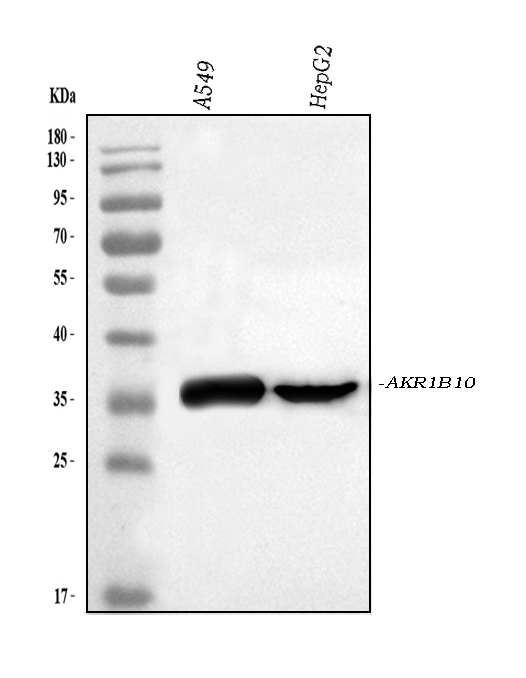

WB
Western blot analysis of AKR1B10 using anti-AKR1B10 antibody. The sample well of each lane was loaded with 30 ug of sample under reducing conditions. Lane 1: A549 whole cell lysates, Lane 2: HepG2 whole cell lysates. After electrophoresis, proteins were transferred to a membrane. Then the membrane was incubated with rabbit anti-AKR1B10 antigen affinity purified polyclonal antibody at a dilution of 1:1000 and probed with a goat anti-rabbit IgG-HRP secondary antibody. The signal is developed using ECL Plus Western Blotting Substrate.IHC
IHC analysis of AKR1B10 using anti-AKR1B10 antibody. AKR1B10 was detected in a paraffin-embedded section of human liver cancer tissue. Biotinylated goat anti-rabbit IgG was used as secondary antibody. The tissue section was incubated with rabbit anti-AKR1B10 Antibody at a dilution of 1:200 and developed using Strepavidin-Biotin-Complex (SABC)with DAB as the chromogen.ICC/IF
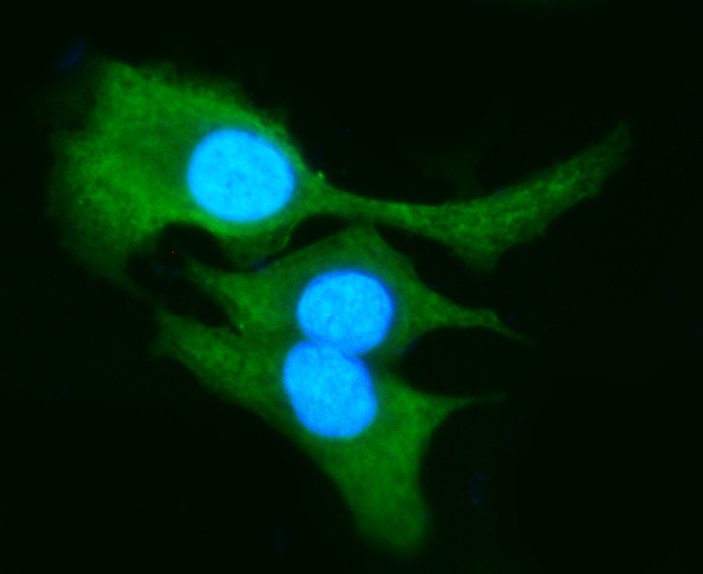
IF analysis of AKR1B10 using anti-AKR1B10 antibody. AKR1B10 was detected in an immunocytochemical section of A549 cells. The section was incubated with rabbit anti-AKR1B10 Antibody at a dilution of 1:100. DyLight®488 Conjugated Goat Anti-Rabbit IgG (Green) was used as secondary antibody. The section was counterstained with DAPI.| Product Name | Rabbit polyclonal antibody to AKR1B10 |
|---|---|
| Antibody Type | Primary Antibodies |
| Immunogen | A synthetic peptide corresponding to a sequence at the C-terminus of human AKR1B10. |
| Clonality | polyclonal |
|---|---|
| Isotype | IgG |
| Host Species | Rabbit |
| Tested Applications | ICC/IFIHCWB |
| WB:1:500-1:2000 IHC:1:50-1:400 ICC/IF:1:50-1:400 |
|
| Species Reactivity | Human |
| Concentration | 0.5mg/ml |
| Purification | Affinity purified |
| Gene Symbol | AKR1B10 |
|---|---|
| Gene Synonyms | HIS HSI ARL1 ARL-1 ALDRLn AKR1B11 AKR1B12 |
| Gene Full Name | aldo-keto reductase family 1 member B10 |
| Gene Summary | This gene encodes a member of the aldo/keto reductase superfamily, which consists of more than 40 known enzymes and proteins. This member can efficiently reduce aliphatic and aromatic aldehydes, and it is less active on hexoses. It is highly expressed in adrenal gland, small intestine, and colon, and may play an important role in liver carcinogenesis. [provided by RefSeq, Jul 2008] |
| Molecular Weight(MW) | 36kDa |
| Cellular Localization | Lysosome,Secreted. |
WB
Western blot analysis of AKR1B10 using anti-AKR1B10 antibody. The sample well of each lane was loaded with 30 ug of sample under reducing conditions. Lane 1: A549 whole cell lysates, Lane 2: HepG2 whole cell lysates. After electrophoresis, proteins were transferred to a membrane. Then the membrane was incubated with rabbit anti-AKR1B10 antigen affinity purified polyclonal antibody at a dilution of 1:1000 and probed with a goat anti-rabbit IgG-HRP secondary antibody. The signal is developed using ECL Plus Western Blotting Substrate.
IHC
IHC analysis of AKR1B10 using anti-AKR1B10 antibody. AKR1B10 was detected in a paraffin-embedded section of human liver cancer tissue. Biotinylated goat anti-rabbit IgG was used as secondary antibody. The tissue section was incubated with rabbit anti-AKR1B10 Antibody at a dilution of 1:200 and developed using Strepavidin-Biotin-Complex (SABC)with DAB as the chromogen.
ICC/IF
IF analysis of AKR1B10 using anti-AKR1B10 antibody. AKR1B10 was detected in an immunocytochemical section of A549 cells. The section was incubated with rabbit anti-AKR1B10 Antibody at a dilution of 1:100. DyLight®488 Conjugated Goat Anti-Rabbit IgG (Green) was used as secondary antibody. The section was counterstained with DAPI.| Application Notes | WB:1:500-1:2000 IHC:1:50-1:400 ICC/IF:1:50-1:400 |
|---|
| Form | Liquid |
|---|---|
| Storage Instructions | 12 months from date of receipt, -20℃ as supplied. 6 months 2 to 8℃ after reconstitution. Avoid repeated freezing and thawing. |
| Storage Buffer | 500ug/ml antibody with PBS, 0.02% NaN3, 1 mg/ml BSA and 50% glycerol. |


 Data sheet for OM643931