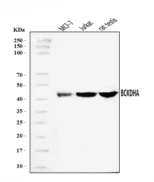
WB
Western blot analysis of BCKDHA using anti-BCKDHA antibody. The sample well of each lane was loaded with 30 ug of sample under reducing conditions. Lane 1: MCF-7 whole cell lysates, Lane 2: Jurkat whole cell lysates, Lane 3: rat testis whole cell lysates. After electrophoresis, proteins were transferred to a membrane. Then the membrane was incubated with rabbit anti-BCKDHA antigen affinity purified polyclonal antibody at a dilution of 1:1000 and probed with a goat anti-rabbit IgG-HRP secondary antibody. The signal is developed using ECL Plus Western Blotting Substrate.IHC
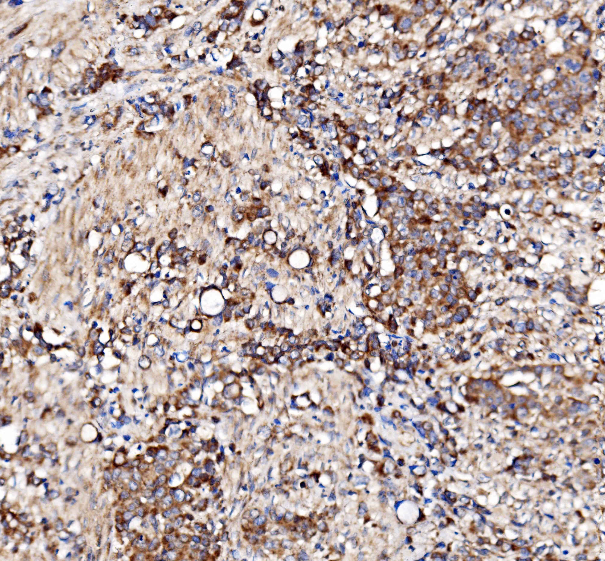
IHC analysis of BCKDHA using anti-BCKDHA antibody. BCKDHA was detected in a paraffin-embedded section of human stomach cancer tissue. Biotinylated goat anti-rabbit IgG was used as secondary antibody. The tissue section was incubated with rabbit anti-BCKDHA Antibody at a dilution of 1:200 and developed using Strepavidin-Biotin-Complex (SABC) with DAB as the chromogen.ICC/IF
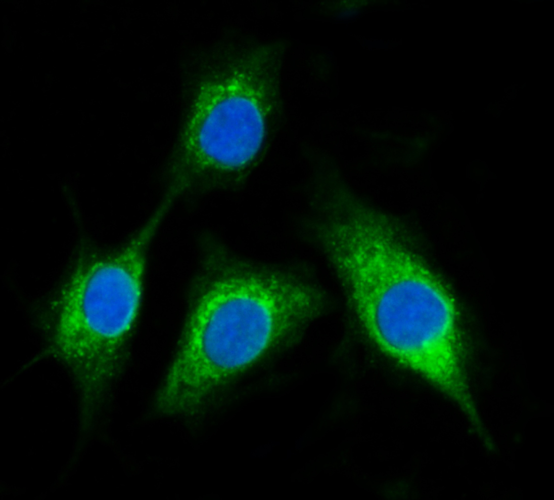
IF analysis of BCKDHA using anti-BCKDHA antibody. BCKDHA was detected in an immunocytochemical section of A549 cells. The section was incubated with rabbit anti-BCKDHA Antibody at a dilution of 1:100. 488 Conjugated Goat Anti-Rabbit IgG (Green) was used as secondary antibody. The section was counterstained with DAP (Blue).FC
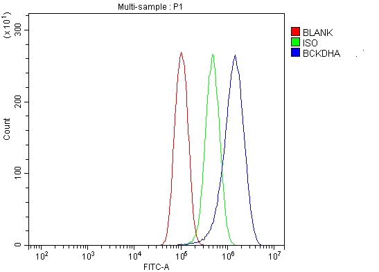
Flow Cytometry analysis of PC-3 cells using anti-BCKDHA antibody. Overlay histogram showing PC-3 cells stained with anti-BCKDHA antibody (Blue line). To facilitate intracellular staining, cells were fixed with 4% paraformaldehyde and permeabilized with permeabilization buffer. The cells were blocked with 10% normal goat serum. And then incubated with rabbit anti-BCKDHA Antibody at 1:100 dilution for 30 min at 20°C. 488 conjugated goat anti-rabbit IgG was used as secondary antibody at 1:100 dilution for 30 minutes at 20°C. Isotype control antibody (Green line) was rabbit IgG at 1:100 dilution used under the same conditions. Unlabelled sample without incubation with primary antibody and secondary antibody (Red line) was used as a blank control.| Product Name | Rabbit polyclonal antibody to BCKDHA |
|---|---|
| Antibody Type | Primary Antibodies |
| Immunogen | E.coli-derived human BCKDHA recombinant protein (Position: A30-K445). |
| Clonality | polyclonal |
|---|---|
| Isotype | IgG |
| Host Species | Rabbit |
| Tested Applications | FCICC/IFIHCWB |
| WB:1:500-1:2000 IHC:1:50-1:400 ICC/IF:1:50-1:400 FC:1:50-1:200 |
|
| Species Reactivity | HumanMouseRat |
| Concentration | 0.5mg/ml |
| Purification | Affinity purified |
| Gene Symbol | BCKDHA |
|---|---|
| Gene Synonyms | MSU MSUD1 OVD1A MSUD1A BCKDE1A |
| Gene Full Name | branched chain keto acid dehydrogenase E1 subunit alpha |
| Gene Summary | The branched-chain alpha-keto acid (BCAA) dehydrogenase (BCKD) complex is an innter mitochondrial enzyme complex that catalyzes the second major step in the catabolism of the branched-chain amino acids leucine, isoleucine, and valine. The BCKD complex consists of three catalytic components: a heterotetrameric (alpha2-beta2) branched-chain alpha-keto acid decarboxylase (E1), a dihydrolipoyl transacylase (E2), and a dihydrolipoamide dehydrogenase (E3). This gene encodes the alpha subunit of the decarboxylase (E1) component. Mutations in this gene result in maple syrup urine disease, type IA. Multiple transcript variants encoding different isoforms have been found for this gene.[provided by RefSeq, Sep 2009] |
| Molecular Weight(MW) | 50kDa |
| Cellular Localization | Mitochondrion matrix. |
WB
Western blot analysis of BCKDHA using anti-BCKDHA antibody. The sample well of each lane was loaded with 30 ug of sample under reducing conditions. Lane 1: MCF-7 whole cell lysates, Lane 2: Jurkat whole cell lysates, Lane 3: rat testis whole cell lysates. After electrophoresis, proteins were transferred to a membrane. Then the membrane was incubated with rabbit anti-BCKDHA antigen affinity purified polyclonal antibody at a dilution of 1:1000 and probed with a goat anti-rabbit IgG-HRP secondary antibody. The signal is developed using ECL Plus Western Blotting Substrate.
IHC
IHC analysis of BCKDHA using anti-BCKDHA antibody. BCKDHA was detected in a paraffin-embedded section of human stomach cancer tissue. Biotinylated goat anti-rabbit IgG was used as secondary antibody. The tissue section was incubated with rabbit anti-BCKDHA Antibody at a dilution of 1:200 and developed using Strepavidin-Biotin-Complex (SABC) with DAB as the chromogen.
ICC/IF
IF analysis of BCKDHA using anti-BCKDHA antibody. BCKDHA was detected in an immunocytochemical section of A549 cells. The section was incubated with rabbit anti-BCKDHA Antibody at a dilution of 1:100. 488 Conjugated Goat Anti-Rabbit IgG (Green) was used as secondary antibody. The section was counterstained with DAP (Blue).
FC
Flow Cytometry analysis of PC-3 cells using anti-BCKDHA antibody. Overlay histogram showing PC-3 cells stained with anti-BCKDHA antibody (Blue line). To facilitate intracellular staining, cells were fixed with 4% paraformaldehyde and permeabilized with permeabilization buffer. The cells were blocked with 10% normal goat serum. And then incubated with rabbit anti-BCKDHA Antibody at 1:100 dilution for 30 min at 20°C. 488 conjugated goat anti-rabbit IgG was used as secondary antibody at 1:100 dilution for 30 minutes at 20°C. Isotype control antibody (Green line) was rabbit IgG at 1:100 dilution used under the same conditions. Unlabelled sample without incubation with primary antibody and secondary antibody (Red line) was used as a blank control.| Application Notes | WB:1:500-1:2000 IHC:1:50-1:400 ICC/IF:1:50-1:400 FC:1:50-1:200 |
|---|
| Form | Liquid |
|---|---|
| Storage Instructions | 12 months from date of receipt, -20℃ as supplied. 6 months 2 to 8℃ after reconstitution. Avoid repeated freezing and thawing. |
| Storage Buffer | 500ug/ml antibody with PBS, 0.02% NaN3, 1 mg/ml BSA and 50% glycerol. |


 Data sheet for OM643938