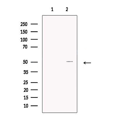

WB
Western blot analysis of extracts from MCF7, using BCKDHA Antibody. The lane on the left was treated with blocking peptide.IHC
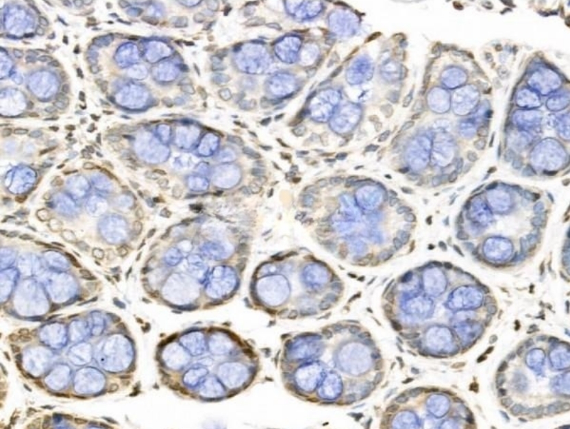
BCKDHA Antibody at 1/100 staining Rat colorectal tissue by IHC-P. The sample was formaldehyde fixed and a heat mediated antigen retrieval step in citrate buffer was performed. The sample was then blocked and incubated with the primary antibody at 4°C overnight. An HRP conjugated anti-Rabbit antibody was used as the secondary antibody.ICC/IF
BCKDHA Antibody staining Hela cells by IF/ICC. The samples were fixed with PFA and permeabilized in 0.1% Triton X-100,then blocked in 10% serum for 45 minutes at 25°C. Samples were then incubated with primary Ab(1:200) and mouse anti-beta tubulin Ab(1:200) for 1 hour at 37°C. An AlexaFluor594 conjugated goat anti-rabbit IgG(H+L) Ab(Red) and an AlexaFluor488 conjugated goat anti-mouse IgG(H+L) Ab(Green) were used as the secondary antibody. The nuclear counter stain is DAPI(blue).| Product Name | Rabbit polyclonal antibody to BCKDHA |
|---|---|
| Antibody Type | Primary Antibodies |
| Immunogen | A synthesized peptide derived from human BCKDHA(Accession P12694), corresponding to amino acid residues A254-F304. |
| Clonality | polyclonal |
|---|---|
| Isotype | IgG |
| Host Species | Rabbit |
| Tested Applications | ICC/IFIHCWB |
| WB:1:500-1:2000 IHC:1:50-1:200 ICC/IF:1:100-1:500 |
|
| Species Reactivity | HumanMouseRat |
| Concentration | 1mg/ml |
| Purification | Affinity purified |
| Gene Symbol | BCKDHA |
|---|---|
| Gene Synonyms | MSU MSUD1 OVD1A MSUD1A BCKDE1A |
| Gene Full Name | branched chain keto acid dehydrogenase E1 subunit alpha |
| Gene Summary | The branched-chain alpha-keto acid (BCAA) dehydrogenase (BCKD) complex is an innter mitochondrial enzyme complex that catalyzes the second major step in the catabolism of the branched-chain amino acids leucine, isoleucine, and valine. The BCKD complex consists of three catalytic components: a heterotetrameric (alpha2-beta2) branched-chain alpha-keto acid decarboxylase (E1), a dihydrolipoyl transacylase (E2), and a dihydrolipoamide dehydrogenase (E3). This gene encodes the alpha subunit of the decarboxylase (E1) component. Mutations in this gene result in maple syrup urine disease, type IA. Multiple transcript variants encoding different isoforms have been found for this gene.[provided by RefSeq, Sep 2009] |
| Molecular Weight(MW) | 50kDa |
| Cellular Localization | Mitochondrion matrix. |
WB
Western blot analysis of extracts from MCF7, using BCKDHA Antibody. The lane on the left was treated with blocking peptide.
IHC
BCKDHA Antibody at 1/100 staining Rat colorectal tissue by IHC-P. The sample was formaldehyde fixed and a heat mediated antigen retrieval step in citrate buffer was performed. The sample was then blocked and incubated with the primary antibody at 4°C overnight. An HRP conjugated anti-Rabbit antibody was used as the secondary antibody.
ICC/IF
BCKDHA Antibody staining Hela cells by IF/ICC. The samples were fixed with PFA and permeabilized in 0.1% Triton X-100,then blocked in 10% serum for 45 minutes at 25°C. Samples were then incubated with primary Ab(1:200) and mouse anti-beta tubulin Ab(1:200) for 1 hour at 37°C. An AlexaFluor594 conjugated goat anti-rabbit IgG(H+L) Ab(Red) and an AlexaFluor488 conjugated goat anti-mouse IgG(H+L) Ab(Green) were used as the secondary antibody. The nuclear counter stain is DAPI(blue).| Application Notes | WB:1:500-1:2000 IHC:1:50-1:200 ICC/IF:1:100-1:500 |
|---|
| Form | Liquid |
|---|---|
| Storage Instructions | Store at -20 °C. Stable for 12 months from date of receipt. |
| Storage Buffer | Rabbit IgG in phosphate buffered saline , pH 7.4, 150mM NaCl, 0.02% sodium azide and 50% glycerol. |


 Data sheet for OM643940