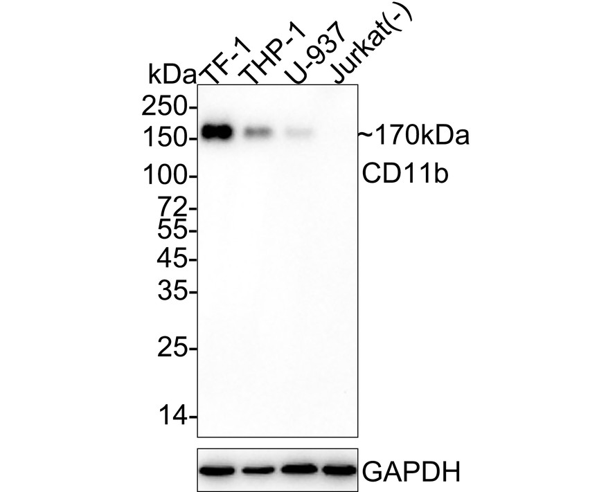
WB
Western blot analysis of CD11b on different lysates with Rabbit anti-CD11b antibody at 1/1,000 dilution. Lane 1: TF-1 cell lysate (10 µg/Lane), Lane 2: THP-1 cell lysate (15 µg/Lane), Lane 3: U-937 cell lysate (30 µg/Lane), Lane 4: Jurkat cell lysate (negative) (10 µg/Lane), Exposure time: 1 minute 50 seconds; 4-20% SDS-PAGE gel. Proteins were transferred to a PVDF membrane and blocked with 5% NFDM/TBST for 1 hour at room temperature. The primary antibody at 1/1,000 dilution was used in 5% NFDM/TBST at 4℃ overnight. Goat Anti-Rabbit IgG - HRP Secondary Antibody at 1/50,000 dilution was used for 1 hour at room temperature.IHC
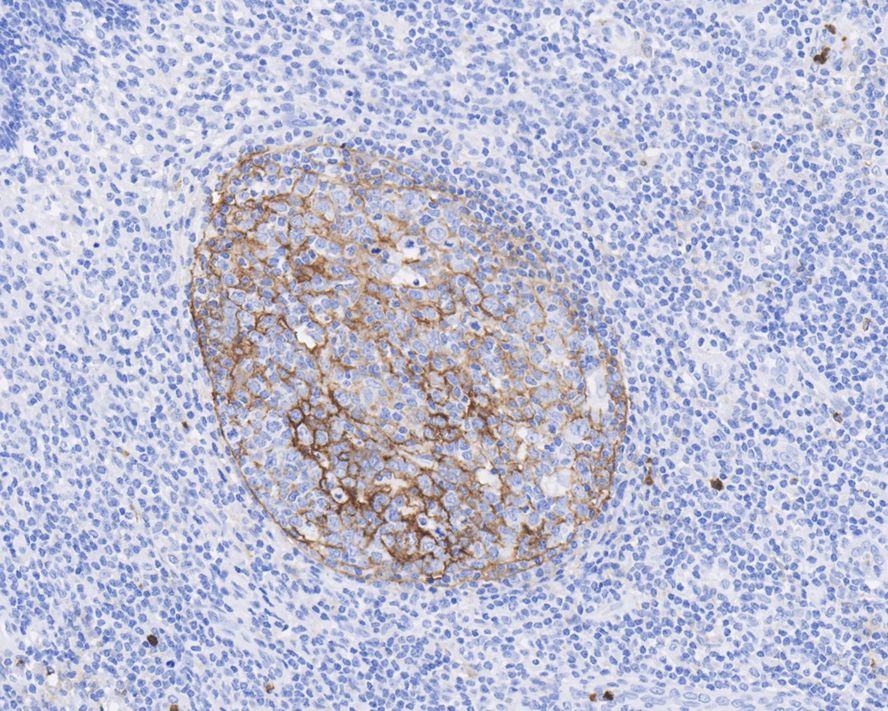
Immunohistochemical analysis of paraffin-embedded human tonsil tissue with Rabbit anti-CD11b antibody at 1/1,000 dilution. The section was pre-treated using heat mediated antigen retrieval with Tris-EDTA buffer (pH 9.0) for 20 minutes. The tissues were blocked in 1% BSA for 20 minutes at room temperature, washed with ddH2O and PBS, and then probed with the primary antibody at 1/1,000 dilution for 1 hour at room temperature. The detection was performed using an HRP conjugated compact polymer system. DAB was used as the chromogen. Tissues were counterstained with hematoxylin and mounted with DPX.ICC/IF
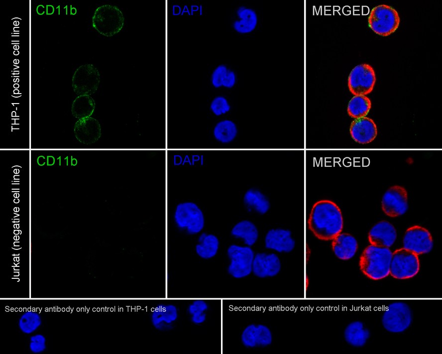
Immunocytochemistry analysis of THP-1 (positive) and Jurkat (negative) labeling CD11b with Rabbit anti-CD11b antibody at 1/100 dilution. Cells were fixed in 100% precooled methanol for 5 minutes at room temperature, then blocked with 1% BSA in 10% negative goat serum for 1 hour at room temperature. Cells were then incubated with Rabbit anti-CD11b antibody at 1/100 dilution in 1% BSA in PBST overnight at 4 ℃. Goat Anti-Rabbit IgG H&L (iFluor™ 488) was used as the secondary antibody at 1/1,000 dilution. PBS instead of the primary antibody was used as the secondary antibody only control. Nuclear DNA was labelled in blue with DAPI. Beta tubulin (red) was stained at 1/100 dilution overnight at +4℃. Goat Anti-Mouse IgG H&L (iFluor™ 594) was used as the secondary antibody at 1/1,000 dilution.IF-P
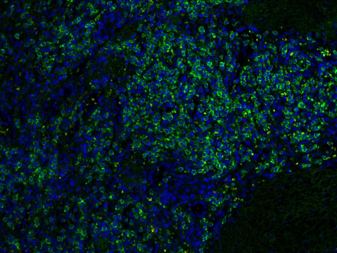
Immunofluorescence analysis of paraffin-embedded human spleen tissue labelling CD11 b. The human spleen section was pre-treated using heat mediated antigen retrieval with Tris-EDTA buffer (pH 9.0) for 20 minutes, blocked in 10% goat serum, and then incubated with at 1/50 dilution , followed by iFluorTM 488 Conjugated Goat anti-rabbit IgG at 1:1000 dilution. Nuclear was stained with Hoechst 33258 at 1/5,000 dilution. Confocal images shows specific membrane staining of CD11b in human spleen.| Product Name | CD11b Recombinant Rabbit Monoclonal Antibody |
|---|---|
| Antibody Type | Primary Antibodies |
| Immunogen | Synthetic peptide within Human CD11b 1103-1152 / 1152. |
| Clonality | monoclonal |
|---|---|
| Isotype | IgG |
| Host Species | Rabbit |
| Tested Applications | ICC/IFIF-PIHCWB |
| WB:1:1000 IHC:1:200-1:1000 ICC/IF:1:50-1:200 IF-P:1:50-1:200 |
|
| Species Reactivity | Human |
| Concentration | 1mg/ml |
| Purification | Protein A |
| Gene Symbol | ITGAM |
|---|---|
| Gene Synonyms | CR3A MO1A CD11B MAC-1 MAC1A SLEB6 |
| Gene Full Name | integrin subunit alpha M |
| Gene Summary | This gene encodes the integrin alpha M chain. Integrins are heterodimeric integral membrane proteins composed of an alpha chain and a beta chain. This I-domain containing alpha integrin combines with the beta 2 chain (ITGB2) to form a leukocyte-specific integrin referred to as macrophage receptor 1 ('Mac-1'), or inactivated-C3b (iC3b) receptor 3 ('CR3'). The alpha M beta 2 integrin is important in the adherence of neutrophils and monocytes to stimulated endothelium, and also in the phagocytosis of complement coated particles. Multiple transcript variants encoding different isoforms have been found for this gene. [provided by RefSeq, Mar 2009] |
| Molecular Weight(MW) | 127kDa(Observed band size: 170kDa) |
| Cellular Localization | Cell membrane, Membrane raft. |
WB
Western blot analysis of CD11b on different lysates with Rabbit anti-CD11b antibody at 1/1,000 dilution. Lane 1: TF-1 cell lysate (10 µg/Lane), Lane 2: THP-1 cell lysate (15 µg/Lane), Lane 3: U-937 cell lysate (30 µg/Lane), Lane 4: Jurkat cell lysate (negative) (10 µg/Lane), Exposure time: 1 minute 50 seconds; 4-20% SDS-PAGE gel. Proteins were transferred to a PVDF membrane and blocked with 5% NFDM/TBST for 1 hour at room temperature. The primary antibody at 1/1,000 dilution was used in 5% NFDM/TBST at 4℃ overnight. Goat Anti-Rabbit IgG - HRP Secondary Antibody at 1/50,000 dilution was used for 1 hour at room temperature.
IHC
Immunohistochemical analysis of paraffin-embedded human tonsil tissue with Rabbit anti-CD11b antibody at 1/1,000 dilution. The section was pre-treated using heat mediated antigen retrieval with Tris-EDTA buffer (pH 9.0) for 20 minutes. The tissues were blocked in 1% BSA for 20 minutes at room temperature, washed with ddH2O and PBS, and then probed with the primary antibody at 1/1,000 dilution for 1 hour at room temperature. The detection was performed using an HRP conjugated compact polymer system. DAB was used as the chromogen. Tissues were counterstained with hematoxylin and mounted with DPX.
ICC/IF
Immunocytochemistry analysis of THP-1 (positive) and Jurkat (negative) labeling CD11b with Rabbit anti-CD11b antibody at 1/100 dilution. Cells were fixed in 100% precooled methanol for 5 minutes at room temperature, then blocked with 1% BSA in 10% negative goat serum for 1 hour at room temperature. Cells were then incubated with Rabbit anti-CD11b antibody at 1/100 dilution in 1% BSA in PBST overnight at 4 ℃. Goat Anti-Rabbit IgG H&L (iFluor™ 488) was used as the secondary antibody at 1/1,000 dilution. PBS instead of the primary antibody was used as the secondary antibody only control. Nuclear DNA was labelled in blue with DAPI. Beta tubulin (red) was stained at 1/100 dilution overnight at +4℃. Goat Anti-Mouse IgG H&L (iFluor™ 594) was used as the secondary antibody at 1/1,000 dilution.
IF-P
Immunofluorescence analysis of paraffin-embedded human spleen tissue labelling CD11 b. The human spleen section was pre-treated using heat mediated antigen retrieval with Tris-EDTA buffer (pH 9.0) for 20 minutes, blocked in 10% goat serum, and then incubated with at 1/50 dilution , followed by iFluorTM 488 Conjugated Goat anti-rabbit IgG at 1:1000 dilution. Nuclear was stained with Hoechst 33258 at 1/5,000 dilution. Confocal images shows specific membrane staining of CD11b in human spleen.| Application Notes | WB:1:1000 IHC:1:200-1:1000 ICC/IF:1:50-1:200 IF-P:1:50-1:200 |
|---|
| Form | Liquid |
|---|---|
| Storage Instructions | Store at +4℃ after thawing. Aliquot store at -20℃. Avoid repeated freeze / thaw cycles. |
| Storage Buffer | PBS (pH7.4), 0.1% BSA, 40% Glycerol. Preservative: 0.05% Sodium Azide. |


 Data sheet for OM643947