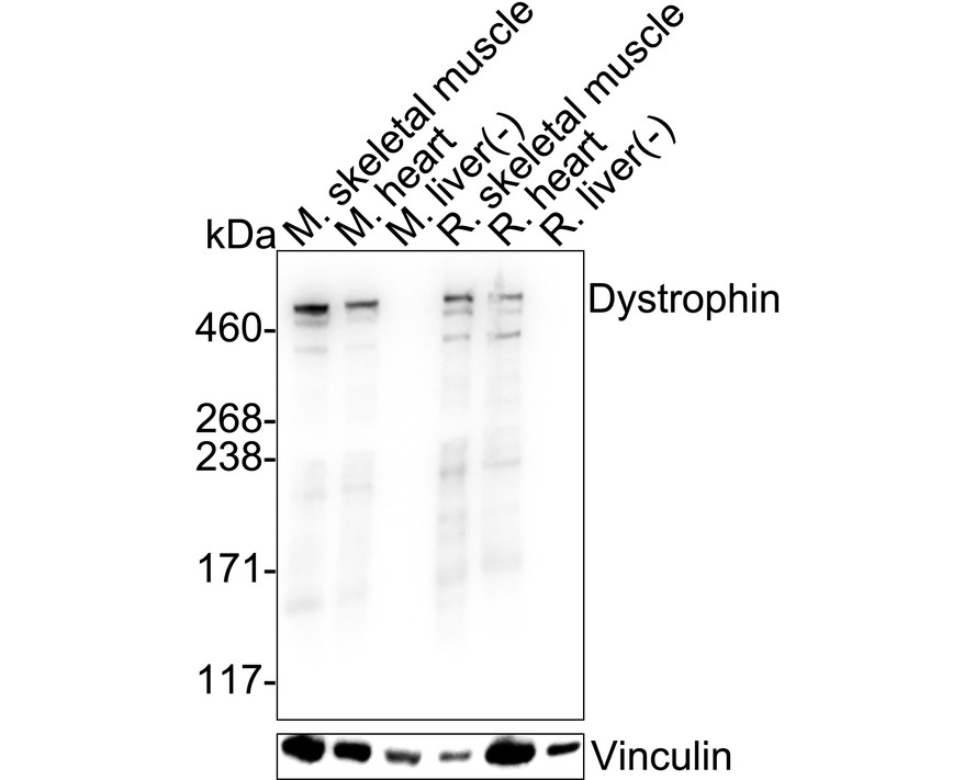
WB
Western blot analysis of Dystrophin on different lysates with Rabbit anti-Dystrophin antibody at 1/1,000 dilution. Lane 1: Mouse skeletal muscle tissue lysate, Lane 2: Mouse heart tissue lysate, Lane 3: Mouse liver tissue lysate (negative), Lane 4: Rat skeletal muscle tissue lysate, Lane 5: Rat heart tissue lysate, Lane 6: Rat liver tissue lysate (negative), Lysates/proteins at 40 µg/Lane. Exposure time: 4 seconds; 3-8% SDS-PAGE gel. Proteins were transferred to a PVDF membrane and blocked with 5% NFDM/TBST for 1 hour at room temperature. The primary antibody at 1/1,000 dilution was used in 5% NFDM/TBST at 4℃ overnight. Goat Anti-Rabbit IgG - HRP Secondary Antibody at 1/50,000 dilution was used for 1 hour at room temperature.IHC
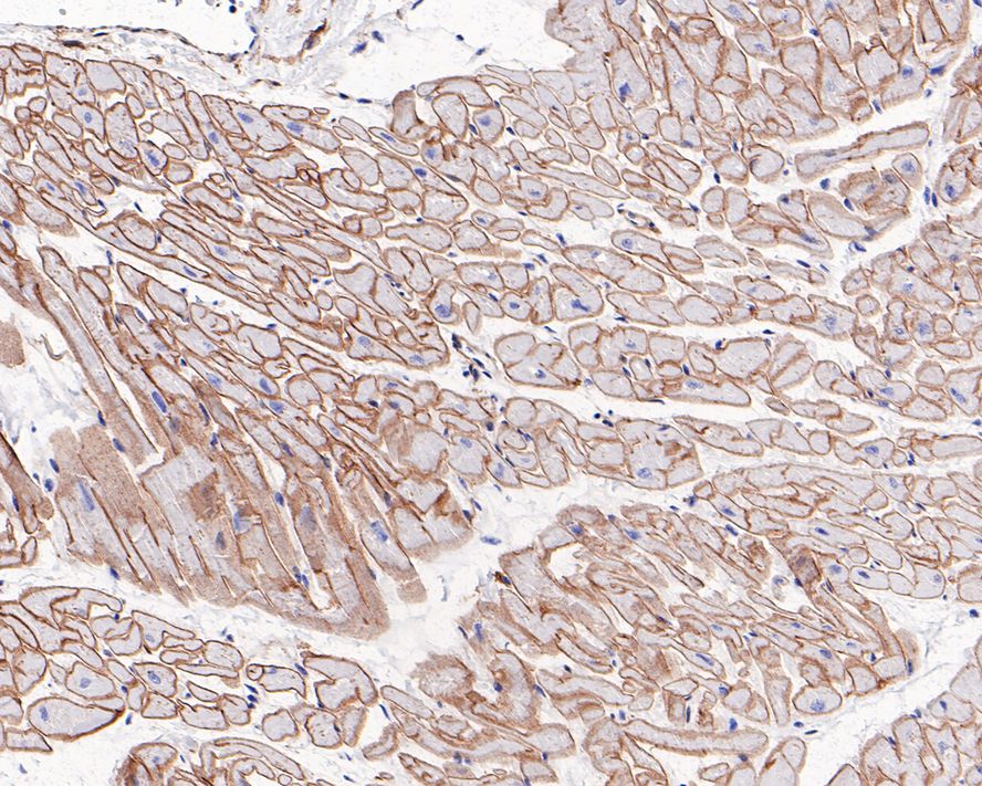
Immunohistochemical analysis of paraffin-embedded human heart tissue with Rabbit anti-Dystrophin antibody at 1/200 dilution. The section was pre-treated using heat mediated antigen retrieval with Tris-EDTA buffer (pH 9.0) for 20 minutes. The tissues were blocked in 1% BSA for 20 minutes at room temperature, washed with ddH2O and PBS, and then probed with the primary antibody at 1/200 dilution for 1 hour at room temperature. The detection was performed using an HRP conjugated compact polymer system. DAB was used as the chromogen. Tissues were counterstained with hematoxylin and mounted with DPX.IHC
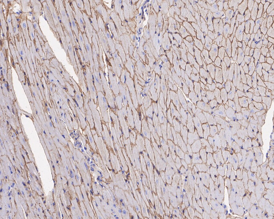
Immunohistochemical analysis of paraffin-embedded mouse heart tissue with Rabbit anti-Dystrophin antibody at 1/1,000 dilution. The section was pre-treated using heat mediated antigen retrieval with sodium citrate buffer (pH 6.0) for 2 minutes. The tissues were blocked in 1% BSA for 20 minutes at room temperature, washed with ddH2O and PBS, and then probed with the primary antibody at 1/1,000 dilution for 1 hour at room temperature. The detection was performed using an HRP conjugated compact polymer system. DAB was used as the chromogen. Tissues were counterstained with hematoxylin and mounted with DPX.IF-F
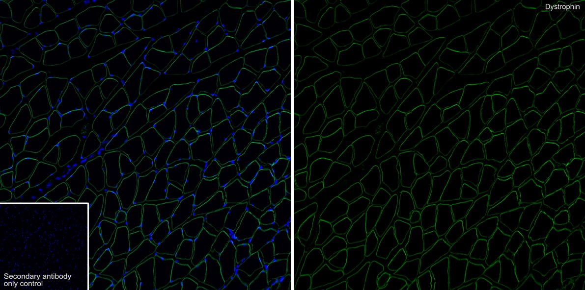
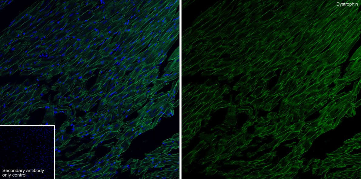
Immunofluorescence analysis of frozen mouse skeletal muscle tissue with Rabbit anti-Dystrophin antibody at 1/200 dilution. The section was pre-treated using heat mediated antigen retrieval with sodium citrate buffer (pH 6.0) for about 2 minutes in microwave oven. The tissues were blocked in 10% negative goat serum for 1 hour at room temperature, washed with PBS, and then probed with the primary antibody (green) at 1/200 dilution overnight at 4 ℃, washed with PBS. Goat Anti-Rabbit IgG H&L (iFluor™ 488) was used as the secondary antibody at 1/1,000 dilution. Nuclei were counterstained with DAPI (blue).IF-F
Immunofluorescence analysis of frozen rat skeletal muscle tissue with Rabbit anti-Dystrophin antibody at 1/200 dilution. The section was pre-treated using heat mediated antigen retrieval with sodium citrate buffer (pH 6.0) for about 2 minutes in microwave oven. The tissues were blocked in 10% negative goat serum for 1 hour at room temperature, washed with PBS, and then probed with the primary antibody (green) at 1/200 dilution overnight at 4 ℃, washed with PBS. Goat Anti-Rabbit IgG H&L (iFluor™ 488) was used as the secondary antibody at 1/1,000 dilution. Nuclei were counterstained with DAPI (blue).| Product Name | Dystrophin Recombinant Rabbit Monoclonal Antibody |
|---|---|
| Antibody Type | Primary Antibodies |
| Immunogen | Synthetic peptide within Human CD11b 1103-1152 / 1152. |
| Clonality | monoclonal |
|---|---|
| Isotype | IgG |
| Host Species | Rabbit |
| Tested Applications | IF-FIHCWB |
| WB:1:1000 IHC:1:200-1:1000 IF-F:1:200 |
|
| Species Reactivity | HumanMouseRat |
| Concentration | 1mg/ml |
| Purification | Protein A |
| Gene Symbol | DMD |
|---|---|
| Gene Synonyms | BMD CMD3B MRX85 DXS142 DXS164 DXS206 DXS230 DXS239 DXS268 DXS269 DXS270 DXS272 |
| Gene Full Name | dystrophin |
| Gene Summary | This gene spans a genomic range of greater than 2 Mb and encodes a large protein containing an N-terminal actin-binding domain and multiple spectrin repeats. The encoded protein forms a component of the dystrophin-glycoprotein complex (DGC), which bridges the inner cytoskeleton and the extracellular matrix. Deletions, duplications, and point mutations at this gene locus may cause Duchenne muscular dystrophy (DMD), Becker muscular dystrophy (BMD), or cardiomyopathy. Alternative promoter usage and alternative splicing result in numerous distinct transcript variants and protein isoforms for this gene. [provided by RefSeq, Dec 2016] |
| Molecular Weight(MW) | 427kDa |
| Cellular Localization | Cell membrane, Cytoplasm, Cell junction. |
WB
Western blot analysis of Dystrophin on different lysates with Rabbit anti-Dystrophin antibody at 1/1,000 dilution. Lane 1: Mouse skeletal muscle tissue lysate, Lane 2: Mouse heart tissue lysate, Lane 3: Mouse liver tissue lysate (negative), Lane 4: Rat skeletal muscle tissue lysate, Lane 5: Rat heart tissue lysate, Lane 6: Rat liver tissue lysate (negative), Lysates/proteins at 40 µg/Lane. Exposure time: 4 seconds; 3-8% SDS-PAGE gel. Proteins were transferred to a PVDF membrane and blocked with 5% NFDM/TBST for 1 hour at room temperature. The primary antibody at 1/1,000 dilution was used in 5% NFDM/TBST at 4℃ overnight. Goat Anti-Rabbit IgG - HRP Secondary Antibody at 1/50,000 dilution was used for 1 hour at room temperature.
IHC
Immunohistochemical analysis of paraffin-embedded human heart tissue with Rabbit anti-Dystrophin antibody at 1/200 dilution. The section was pre-treated using heat mediated antigen retrieval with Tris-EDTA buffer (pH 9.0) for 20 minutes. The tissues were blocked in 1% BSA for 20 minutes at room temperature, washed with ddH2O and PBS, and then probed with the primary antibody at 1/200 dilution for 1 hour at room temperature. The detection was performed using an HRP conjugated compact polymer system. DAB was used as the chromogen. Tissues were counterstained with hematoxylin and mounted with DPX.
IHC
Immunohistochemical analysis of paraffin-embedded mouse heart tissue with Rabbit anti-Dystrophin antibody at 1/1,000 dilution. The section was pre-treated using heat mediated antigen retrieval with sodium citrate buffer (pH 6.0) for 2 minutes. The tissues were blocked in 1% BSA for 20 minutes at room temperature, washed with ddH2O and PBS, and then probed with the primary antibody at 1/1,000 dilution for 1 hour at room temperature. The detection was performed using an HRP conjugated compact polymer system. DAB was used as the chromogen. Tissues were counterstained with hematoxylin and mounted with DPX.
IF-F
Immunofluorescence analysis of frozen mouse skeletal muscle tissue with Rabbit anti-Dystrophin antibody at 1/200 dilution. The section was pre-treated using heat mediated antigen retrieval with sodium citrate buffer (pH 6.0) for about 2 minutes in microwave oven. The tissues were blocked in 10% negative goat serum for 1 hour at room temperature, washed with PBS, and then probed with the primary antibody (green) at 1/200 dilution overnight at 4 ℃, washed with PBS. Goat Anti-Rabbit IgG H&L (iFluor™ 488) was used as the secondary antibody at 1/1,000 dilution. Nuclei were counterstained with DAPI (blue).
IF-F
Immunofluorescence analysis of frozen rat skeletal muscle tissue with Rabbit anti-Dystrophin antibody at 1/200 dilution. The section was pre-treated using heat mediated antigen retrieval with sodium citrate buffer (pH 6.0) for about 2 minutes in microwave oven. The tissues were blocked in 10% negative goat serum for 1 hour at room temperature, washed with PBS, and then probed with the primary antibody (green) at 1/200 dilution overnight at 4 ℃, washed with PBS. Goat Anti-Rabbit IgG H&L (iFluor™ 488) was used as the secondary antibody at 1/1,000 dilution. Nuclei were counterstained with DAPI (blue).| Application Notes | WB:1:1000 IHC:1:200-1:1000 IF-F:1:200 |
|---|
| Form | Liquid |
|---|---|
| Storage Instructions | Store at +4℃ after thawing. Aliquot store at -20℃. Avoid repeated freeze / thaw cycles. |
| Storage Buffer | PBS (pH7.4), 0.1% BSA, 40% Glycerol. Preservative: 0.05% Sodium Azide. |


 Data sheet for OM643958