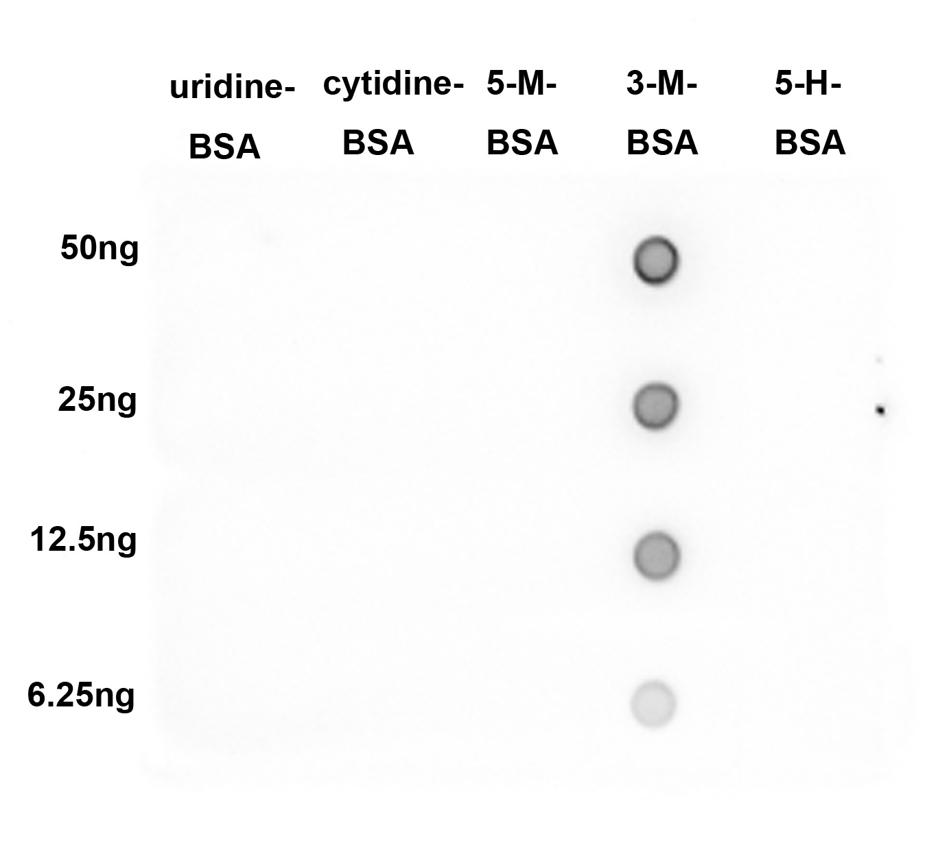
DB
Dot blot analysis of 3mC on different proteins with Rabbit anti-3mC antibody at 1/2,000 dilution. Goat Anti-Rabbit IgG - HRP Secondary Antibody at 1/50,000 dilution for 1 hour at room temperature. Lane 1: Uridine-BSA (negative), Lane 2: Cytidine-BSA (negative), Lane 3: 5-Methylcytosine-BSA (negative), Lane 4: 3-Methylcytosine-BSA (positive), Lane 5: 5-Hydroxymethylcytidine-BSA (negative), Proteins loading: 50ng, 25ng, 12.5ng, 6.25ng; Blocking and dilution buffer: 5% NFDM/TBST; Exposure time: 1 minute.ICC/IF
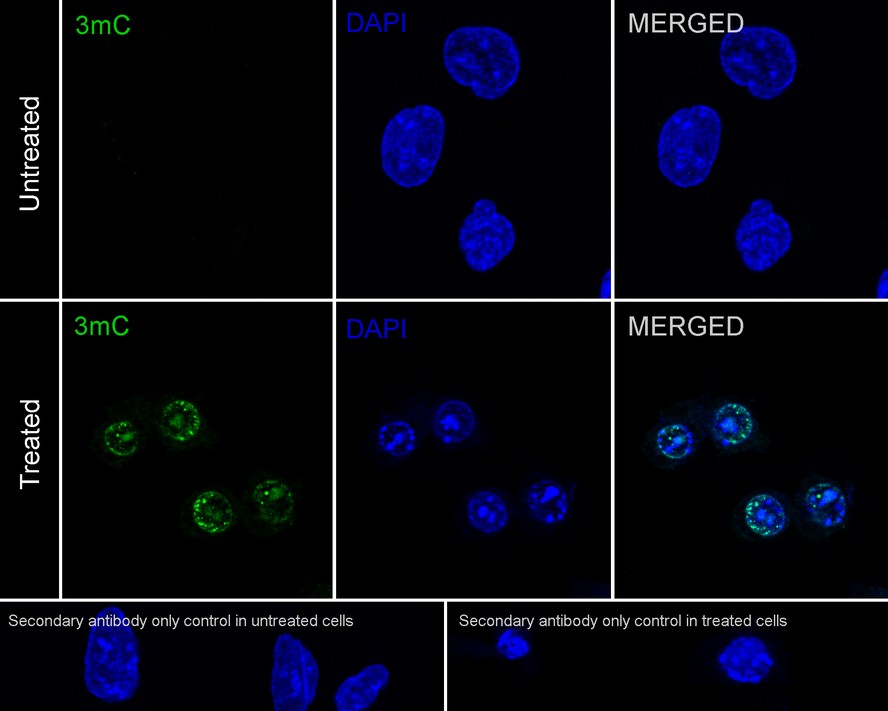
Immunocytochemistry analysis of U-2 OS cells treated with 4mM Methyl Methanesulfonate for 1 hour labeling 3mC with Rabbit anti-3mC antibody at 1/100 dilution. Cells were fixed in 70% ethyl alcohol for 5 minutes at room temperature, then subjected to acid hydrolysis using 2M HCl in TBST for 30 minutes at room temperature. Permeabilized with 0.1% Triton X-100 in PBS for 15 minutes, and then blocked with 2% BSA for 30 minutes at room temperature. Cells were then incubated with Rabbit anti-3mC antibody at 1/100 dilution in 1% BSA in PBST overnight at 4 ℃. Goat Anti-Rabbit IgG H&L (iFluor™ 488) was used as the secondary antibody at 1/1,000 dilution. PBS instead of the primary antibody was used as the secondary antibody only control. Nuclear DNA was labelled in blue with DAPI.| Product Name | 3mC Recombinant Rabbit Monoclonal Antibody |
|---|---|
| Antibody Type | Primary Antibodies |
| Immunogen | 3-mc-OVA |
| Clonality | monoclonal |
|---|---|
| Isotype | IgG |
| Host Species | Rabbit |
| Tested Applications | DBICC/IF |
| DB:1:2000 ICC/IF:1:100 |
|
| Species Reactivity | All |
| Concentration | 1mg/ml |
| Purification | Protein A |
| Molecular Weight(MW) | 12kDa(Observed band size: 17kDa) |
|---|---|
| Function | Methylation of DNA can occur non-enzymatically at the nitrogen-three of the cytosine base through spontaneous exposure to endogenous S-adenosyl methionine (SAM). The resulting 3-methylcytosine (3-mC) is mutagenic and must be repaired, which occurs in humans through the base excision repair (BER) or dealkylation via human homologues of the E. coli AlkB protein. 3-methylcytosine is present in human cell lines and increased levels of 3-mC impair proliferation. |
DB
Dot blot analysis of 3mC on different proteins with Rabbit anti-3mC antibody at 1/2,000 dilution. Goat Anti-Rabbit IgG - HRP Secondary Antibody at 1/50,000 dilution for 1 hour at room temperature. Lane 1: Uridine-BSA (negative), Lane 2: Cytidine-BSA (negative), Lane 3: 5-Methylcytosine-BSA (negative), Lane 4: 3-Methylcytosine-BSA (positive), Lane 5: 5-Hydroxymethylcytidine-BSA (negative), Proteins loading: 50ng, 25ng, 12.5ng, 6.25ng; Blocking and dilution buffer: 5% NFDM/TBST; Exposure time: 1 minute.
ICC/IF
Immunocytochemistry analysis of U-2 OS cells treated with 4mM Methyl Methanesulfonate for 1 hour labeling 3mC with Rabbit anti-3mC antibody at 1/100 dilution. Cells were fixed in 70% ethyl alcohol for 5 minutes at room temperature, then subjected to acid hydrolysis using 2M HCl in TBST for 30 minutes at room temperature. Permeabilized with 0.1% Triton X-100 in PBS for 15 minutes, and then blocked with 2% BSA for 30 minutes at room temperature. Cells were then incubated with Rabbit anti-3mC antibody at 1/100 dilution in 1% BSA in PBST overnight at 4 ℃. Goat Anti-Rabbit IgG H&L (iFluor™ 488) was used as the secondary antibody at 1/1,000 dilution. PBS instead of the primary antibody was used as the secondary antibody only control. Nuclear DNA was labelled in blue with DAPI.| Application Notes | DB:1:2000 ICC/IF:1:100 |
|---|
| Form | Liquid |
|---|---|
| Storage Instructions | Store at +4℃ after thawing. Aliquot store at -20℃. Avoid repeated freeze / thaw cycles. |
| Storage Buffer | 1*TBS (pH7.4), 0.05% BSA, 40% Glycerol. Preservative: 0.05% Sodium Azide. |


 Data sheet for OM643988