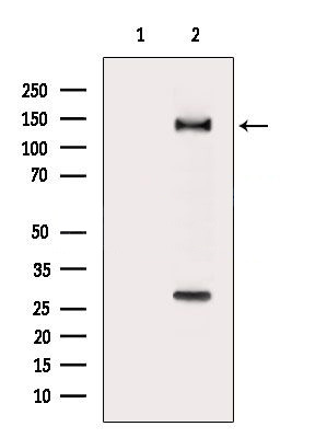
WB
Western blot analysis of extracts from MCF7 cells(serum starvation treatment), using RAPH1 Antibody. The lane on the left was treated with blocking peptide.IHC
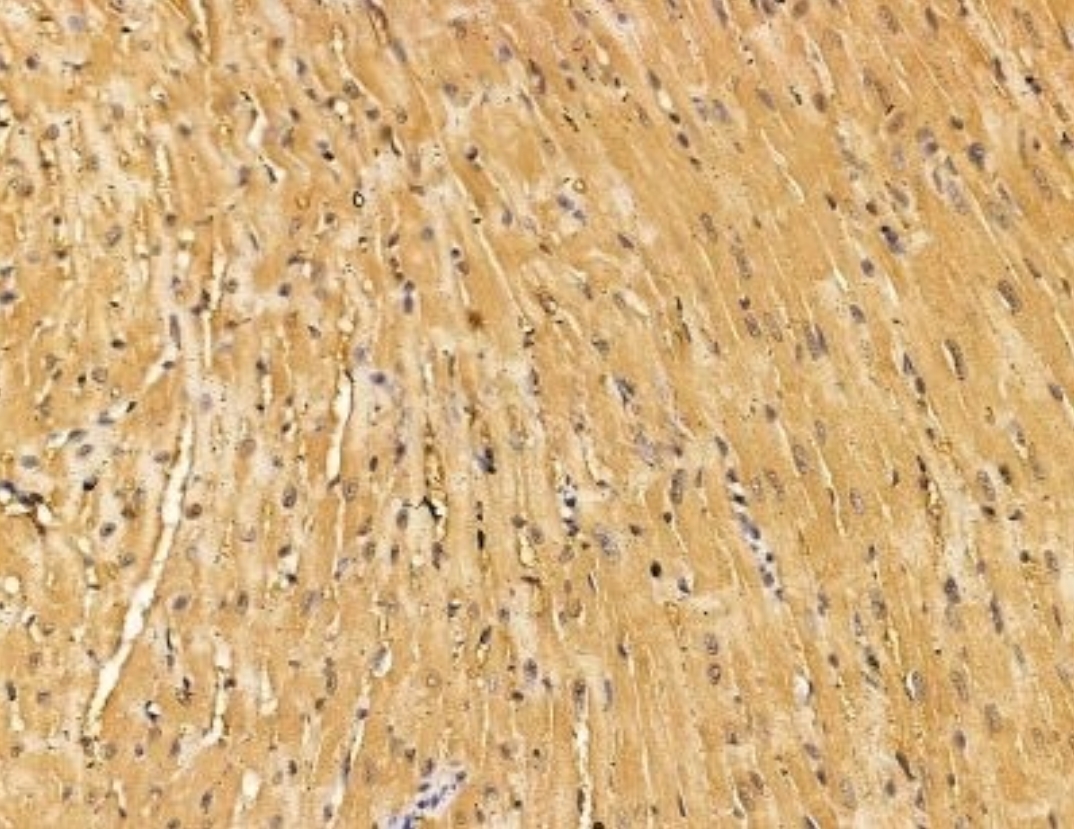
RAPH1 Antibody at 1/100 staining mouse heart tissue by IHC-P. The sample was formaldehyde fixed and a heat mediated antigen retrieval step in citrate buffer was performed. The sample was then blocked and incubated with the primary antibody at 4°C overnight. An HRP conjugated anti-rabbit antibody was used as the secondary antibody.ICC/IF
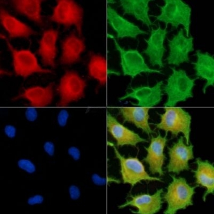
RAPH1 Antibody staining A549 cells by IF/ICC. The samples were fixed with PFA and permeabilized in 0.1% Triton X-100, then blocked in 10% serum for 45 minutes at 25°C. Samples were then incubated with primary Ab and mouse anti-beta tubulin Ab for 1 hour at 37°C. An AlexaFluor594 conjugated goat anti-rabbit IgG Ab(Red) and an AlexaFluor488 conjugated goat anti-mouse IgG Ab(Green) were used as the secondary antibody. The nuclear counter stain is DAPI (blue).| Product Name | RRabbit polyclonal antibody to RAPH1 |
|---|---|
| Antibody Type | Primary Antibodies |
| Clonality | polyclonal |
|---|---|
| Isotype | IgG |
| Host Species | Rabbit |
| Tested Applications | ICC/IFIHCWB |
| WB:1:1000-1:2000 IHC:1:50-1:200 ICC/IF:1:100-1:500 |
|
| Species Reactivity | Human |
| Concentration | 1mg/ml |
| Purification | Affinity purified |
| Gene Symbol | RAPH1 |
|---|---|
| Gene Synonyms | LPD RMO1 PREL2 PREL-2 ALS2CR9 ALS2CR18 RalGDS/AF-6 |
| Gene Full Name | Ras association (RalGDS/AF-6) and pleckstrin homology domains 1 |
| Gene Summary | This gene encodes a protein that belongs to the Mig10/Rap1-interacting adaptor molecule/Lamellipodin family of adapter proteins, which function in cell migration. Members of this family contain pleckstrin-homology domains, Ras-association domains, and proline-rich C-termini. The protein encoded by this gene regulates actin dynamics through interaction with Ena/Vasodilator proteins as well as direct binding to filamentous actin to regulate actin network assembly. Alternative splicing results in multiple transcript variants. [provided by RefSeq, Jul 2016] |
| Molecular Weight(MW) | 135kDa |
| Cellular Localization | Cell membrane,Cytoplasm,Cytoskeleton. |
WB
Western blot analysis of extracts from MCF7 cells(serum starvation treatment), using RAPH1 Antibody. The lane on the left was treated with blocking peptide.
IHC
RAPH1 Antibody at 1/100 staining mouse heart tissue by IHC-P. The sample was formaldehyde fixed and a heat mediated antigen retrieval step in citrate buffer was performed. The sample was then blocked and incubated with the primary antibody at 4°C overnight. An HRP conjugated anti-rabbit antibody was used as the secondary antibody.
ICC/IF
RAPH1 Antibody staining A549 cells by IF/ICC. The samples were fixed with PFA and permeabilized in 0.1% Triton X-100, then blocked in 10% serum for 45 minutes at 25°C. Samples were then incubated with primary Ab and mouse anti-beta tubulin Ab for 1 hour at 37°C. An AlexaFluor594 conjugated goat anti-rabbit IgG Ab(Red) and an AlexaFluor488 conjugated goat anti-mouse IgG Ab(Green) were used as the secondary antibody. The nuclear counter stain is DAPI (blue).| Application Notes | WB:1:1000-1:2000 IHC:1:50-1:200 ICC/IF:1:100-1:500 |
|---|
| Form | Liquid |
|---|---|
| Storage Instructions | Store at -20 °C. Stable for 12 months from date of receipt. |
| Storage Buffer | Rabbit IgG in phosphate buffered saline , pH 7.4, 150mM NaCl, 0.02% sodium azide and 50% glycerol. |


 Data sheet for OM644019