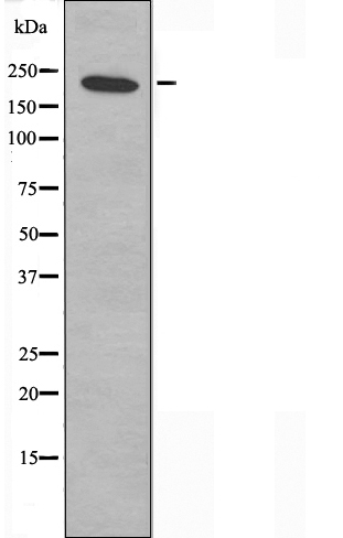
WB
Western blot analysis of extracts from 293 cells, using DIDO1 antibody.IHC
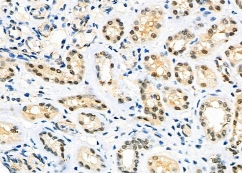
DIDO1 Antibody at 1/100 staining Human kidney cancer and adjacent normal tissues by IHC-P. The sample was formaldehyde fixed and a heat mediated antigen retrieval step in citrate buffer was performed. The sample was then blocked and incubated with the primary antibody at 4°C overnight. An HRP conjugated anti-Rabbit antibody was used as the secondary antibody.ICC/IF
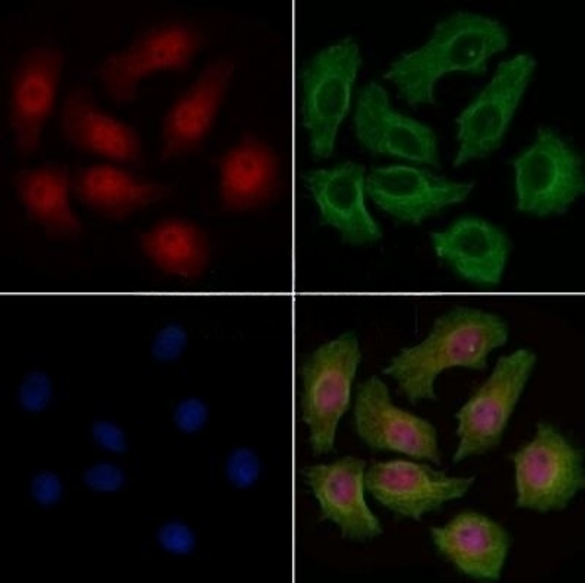
DIDO1 Antibody staining HepG2 cells by IF/ICC. The samples were fixed with PFA and permeabilized in 0.1% Triton X-100,then blocked in 10% serum for 45 minutes at 25°C. Samples were then incubated with primary Ab(1:200) and mouse anti-beta tubulin Ab(1:200) for 1 hour at 37°C. An AlexaFluor594 conjugated goat anti-rabbit IgG(H+L) Ab(Red) and an AlexaFluor488 conjugated goat anti-mouse IgG(H+L) Ab(Green) were used as the secondary antibody. The nuclear counter stain is DAPI(blue).| Product Name | Rabbit polyclonal antibody to DIDO1 |
|---|---|
| Antibody Type | Primary Antibodies |
| Clonality | polyclonal |
|---|---|
| Isotype | IgG |
| Host Species | Rabbit |
| Tested Applications | ICC/IFIHCWB |
| WB:1:500-1:1000 IHC:1:50-1:200 ICC/IF:1:100-1:500 |
|
| Species Reactivity | HumanMouse |
| Concentration | 1mg/ml |
| Purification | Affinity purified |
| Gene Symbol | DIDO1 |
|---|---|
| Gene Synonyms | BYE1 DIO1 DATF1 DIDO2 DIDO3 DIO-1 DATF-1 C20orf158 dJ885L7.8 |
| Gene Full Name | death inducer-obliterator 1 |
| Gene Summary | Apoptosis, a major form of cell death, is an efficient mechanism for eliminating unwanted cells and is of central importance for development and homeostasis in metazoan animals. In mice, the death inducer-obliterator-1 gene is upregulated by apoptotic signals and encodes a cytoplasmic protein that translocates to the nucleus upon apoptotic signal activation. When overexpressed, the mouse protein induced apoptosis in cell lines growing in vitro. This gene is similar to the mouse gene and therefore is thought to be involved in apoptosis. Alternatively spliced transcripts have been found for this gene, encoding multiple isoforms. [provided by RefSeq, Jul 2008] |
| Molecular Weight(MW) | 244kDa |
| Cellular Localization | Cytoplasm,Cytoskeleton,Nucleus. |
WB
Western blot analysis of extracts from 293 cells, using DIDO1 antibody.
IHC
DIDO1 Antibody at 1/100 staining Human kidney cancer and adjacent normal tissues by IHC-P. The sample was formaldehyde fixed and a heat mediated antigen retrieval step in citrate buffer was performed. The sample was then blocked and incubated with the primary antibody at 4°C overnight. An HRP conjugated anti-Rabbit antibody was used as the secondary antibody.
ICC/IF
DIDO1 Antibody staining HepG2 cells by IF/ICC. The samples were fixed with PFA and permeabilized in 0.1% Triton X-100,then blocked in 10% serum for 45 minutes at 25°C. Samples were then incubated with primary Ab(1:200) and mouse anti-beta tubulin Ab(1:200) for 1 hour at 37°C. An AlexaFluor594 conjugated goat anti-rabbit IgG(H+L) Ab(Red) and an AlexaFluor488 conjugated goat anti-mouse IgG(H+L) Ab(Green) were used as the secondary antibody. The nuclear counter stain is DAPI(blue).| Application Notes | WB:1:500-1:1000 IHC:1:50-1:200 ICC/IF:1:100-1:500 |
|---|
| Form | Liquid |
|---|---|
| Storage Instructions | Store at -20 °C. Stable for 12 months from date of receipt. |
| Storage Buffer | Rabbit IgG in phosphate buffered saline , pH 7.4, 150mM NaCl, 0.02% sodium azide and 50% glycerol. |


 Data sheet for OM644021