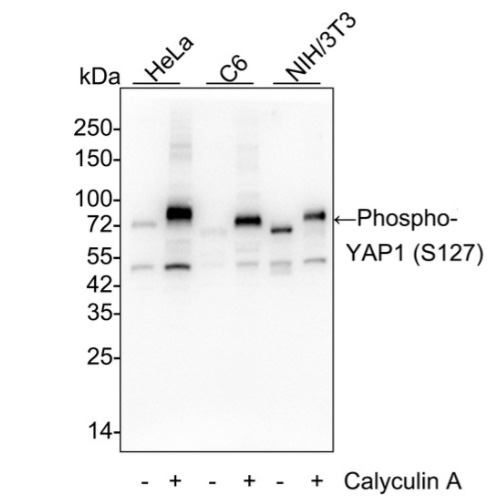
WB
Western blot analysis of Phospho-YAP1 (S127) on different lysates with Rabbit anti-Phospho-YAP1 (S127) antibody at 1/5,000 dilution. Lane 1: HeLa cell lysate, Lane 2: HeLa treated with 100nM Calyculin A for 30 minutes cell lysate, Lane 3: C6 cell lysate, Lane 4: C6 treated with 100nM Calyculin A for 30 minutes cell lysate, Lane 5: NIH/3T3 cell lysate, Lane 6: NIH/3T3 starved for 24 hours then treated with 100nM Calyculin A for 30 minutes cell lysate, Lysates/proteins at 20 µg/Lane. Exposure time: 50 seconds; 4-20% SDS-PAGE gel. Proteins were transferred to a PVDF membrane and blocked with 5% NFDM/TBST for 1 hour at room temperature. The primary antibody at 1/5,000 dilution was used in 5% BSA at 4℃ overnight. Goat Anti-Rabbit IgG - HRP Secondary Antibody at 1/50,000 dilution was used for 1 hour at room temperature.IHC
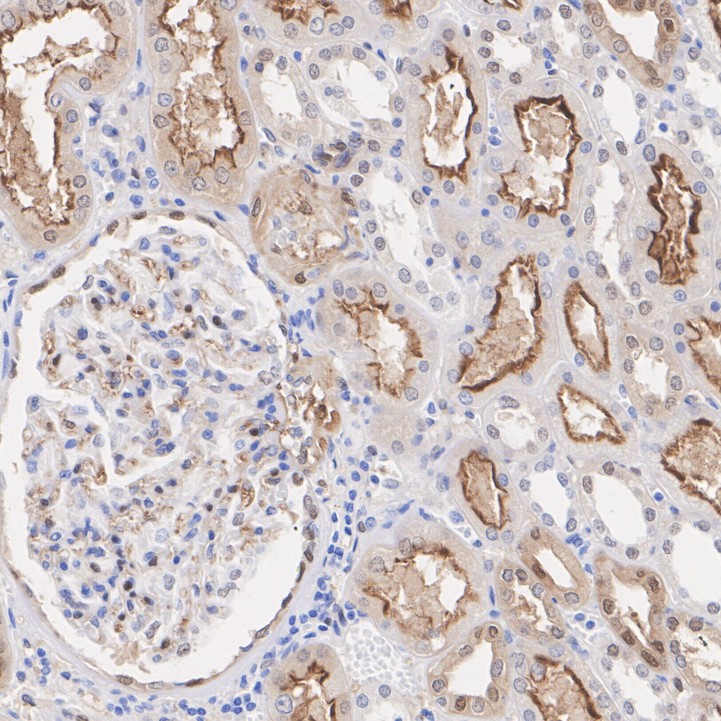
Immunohistochemical analysis of paraffin-embedded human kidney tissue with Rabbit anti-Phospho-YAP1 (S127) antibody at 1/5,000 dilution. The secion was tpre-treated using heat mediated antigen retrieval with Tris-EDTA buffer (pH 9.0) for 20 minutes. The tissues were blocked in 1% BSA for 20 minutes at room temperature, washed with ddH2O and PBS, and then probed with the primary antibody at 1/5,000 dilution for 1 hour at room temperature. The detection was performed using an HRP conjugated compact polymer system. DAB was used as the chromogen. Tissues were counterstained with hematoxylin and mounted with DPX.IHC
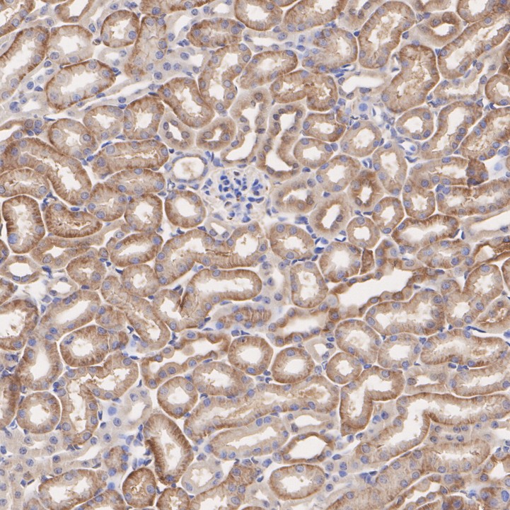
Immunohistochemical analysis of paraffin-embedded mouse kidney tissue with Rabbit anti-Phospho-YAP1 (S127) antibody at 1/5,000 dilution. The section was pre-treated using heat mediated antigen retrieval with Tris-EDTA buffer (pH 9.0) for 20 minutes. The tissues were blocked in 1% BSA for 20 minutes at room temperature, washed with ddH2O and PBS, and then probed with the primary antibody at 1/5,000 dilution for 1 hour at room temperature. The detection was performed using an HRP conjugated compact polymer system. DAB was used as the chromogen. Tissues were counterstained with hematoxylin and mounted with DPX.| Product Name | Phospho-YAP1 (S127) Recombinant Rabbit Monoclonal Antibody |
|---|---|
| Antibody Type | Primary Antibodies |
| Immunogen | Synthetic phospho-peptide corresponding to residues surrounding Ser127 of human YAP1. |
| Clonality | monoclonal |
|---|---|
| Isotype | IgG |
| Host Species | Rabbit |
| Tested Applications | IHCWB |
| WB:1:5000 IHC:1:2000-1:5000 |
|
| Species Reactivity | HumanMouseRat |
| Concentration | 1mg/ml |
| Purification | Protein A |
| Gene Symbol | YAP1 |
|---|---|
| Gene Synonyms | YAP YKI COB1 YAP2 YAP-1 YAP65 |
| Gene Full Name | Yes1 associated transcriptional regulator |
| Gene Summary | This gene encodes a downstream nuclear effector of the Hippo signaling pathway which is involved in development, growth, repair, and homeostasis. This gene is known to play a role in the development and progression of multiple cancers as a transcriptional regulator of this signaling pathway and may function as a potential target for cancer treatment. Alternative splicing results in multiple transcript variants encoding different isoforms. [provided by RefSeq, Aug 2013] |
| Molecular Weight(MW) | 54kDa(Observed band size: 70kDa) |
| Function | The Yes-associated protein, otherwise known as YAP, is a 14-3-3-binding molecule that was originally recognized by virtue of its ability to bind to the SH3 domain of Yes. The binding of YAP to 14-3-3 requires the phosphorylation of a homologous serine residue (Ser 112) in the YAP 14-3-3-binding motif. The highly conserved and ubiquitously expressed 14-3-3 proteins regulate differentiation, cell cycle progression and apoptosis by binding intracellular phosphoproteins involved in signal transduction. YAP may link events at the plasma membrane and cytoskeleton to inhibition of transcription in the nucleus in a manner regulated by 14-3-3 proteins. YAP shares homology with the WW domain of TAZ, transcriptional co-activator with PDZ-binding motif, which functions as a transcriptional co-activator by binding to the PPXY motif present in transcription factors. YAP is expressed at high levels in the lung, placenta, prostate, ovary and testis. |
| Cellular Localization | Cytoplasm, Nucleus. |
WB
Western blot analysis of Phospho-YAP1 (S127) on different lysates with Rabbit anti-Phospho-YAP1 (S127) antibody at 1/5,000 dilution. Lane 1: HeLa cell lysate, Lane 2: HeLa treated with 100nM Calyculin A for 30 minutes cell lysate, Lane 3: C6 cell lysate, Lane 4: C6 treated with 100nM Calyculin A for 30 minutes cell lysate, Lane 5: NIH/3T3 cell lysate, Lane 6: NIH/3T3 starved for 24 hours then treated with 100nM Calyculin A for 30 minutes cell lysate, Lysates/proteins at 20 µg/Lane. Exposure time: 50 seconds; 4-20% SDS-PAGE gel. Proteins were transferred to a PVDF membrane and blocked with 5% NFDM/TBST for 1 hour at room temperature. The primary antibody at 1/5,000 dilution was used in 5% BSA at 4℃ overnight. Goat Anti-Rabbit IgG - HRP Secondary Antibody at 1/50,000 dilution was used for 1 hour at room temperature.
IHC
Immunohistochemical analysis of paraffin-embedded human kidney tissue with Rabbit anti-Phospho-YAP1 (S127) antibody at 1/5,000 dilution. The secion was tpre-treated using heat mediated antigen retrieval with Tris-EDTA buffer (pH 9.0) for 20 minutes. The tissues were blocked in 1% BSA for 20 minutes at room temperature, washed with ddH2O and PBS, and then probed with the primary antibody at 1/5,000 dilution for 1 hour at room temperature. The detection was performed using an HRP conjugated compact polymer system. DAB was used as the chromogen. Tissues were counterstained with hematoxylin and mounted with DPX.
IHC
Immunohistochemical analysis of paraffin-embedded mouse kidney tissue with Rabbit anti-Phospho-YAP1 (S127) antibody at 1/5,000 dilution. The section was pre-treated using heat mediated antigen retrieval with Tris-EDTA buffer (pH 9.0) for 20 minutes. The tissues were blocked in 1% BSA for 20 minutes at room temperature, washed with ddH2O and PBS, and then probed with the primary antibody at 1/5,000 dilution for 1 hour at room temperature. The detection was performed using an HRP conjugated compact polymer system. DAB was used as the chromogen. Tissues were counterstained with hematoxylin and mounted with DPX.| Application Notes | WB:1:5000 IHC:1:2000-1:5000 |
|---|
| Form | Liquid |
|---|---|
| Storage Instructions | Store at +4℃ after thawing. Aliquot store at -20℃. Avoid repeated freeze / thaw cycles. |
| Storage Buffer | 1*TBS (pH7.4), 0.05% BSA, 40% Glycerol. Preservative: 0.05% Sodium Azide. |


 Data sheet for OM644036