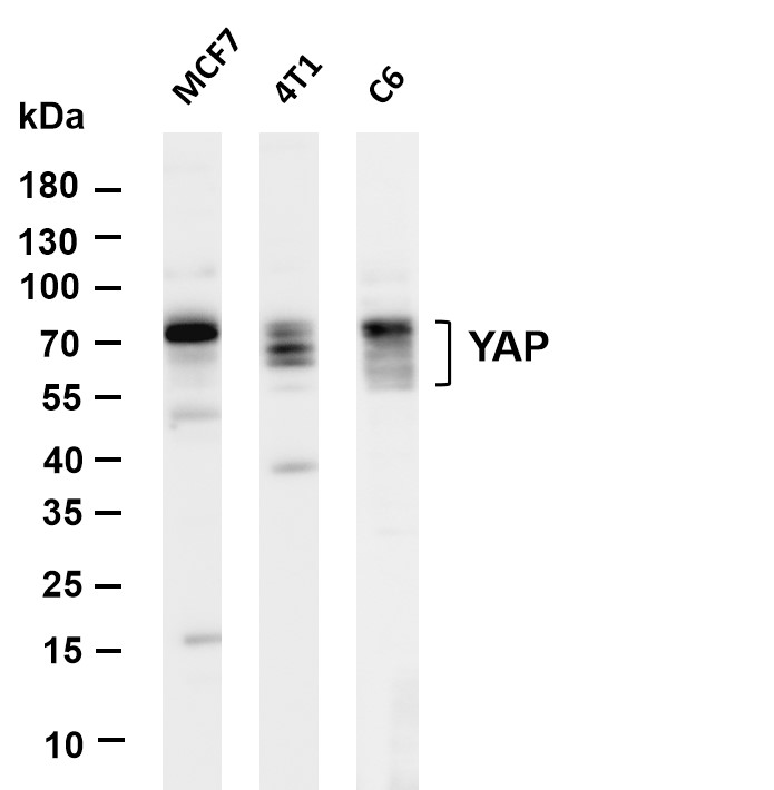
WB
Various whole cell lysates were separated by 4-20% SDS-PAGE, and the membrane was blotted with anti-YAP antibody. The HRP-conjugated Goat anti-Rabbit IgG(H + L) antibody was used to detect the antibody. Lane 1: MCF7, Lane 2: 4T1, Lane 3: C6.IHC
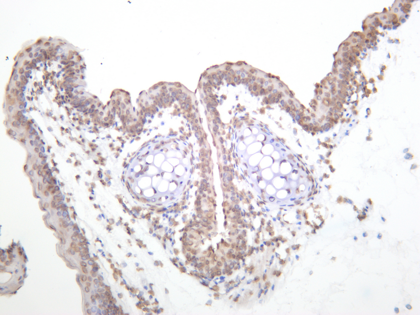
Mouse thyroid was stained with anti-YAP rabbit antibody.IHC
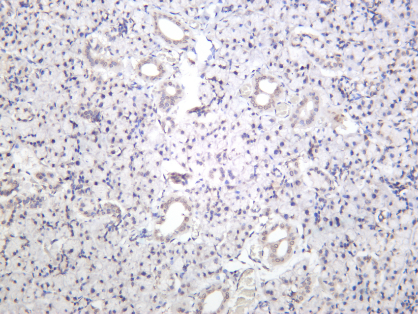
Rat thyroid was stained with anti-YAP rabbit antibody.IHC
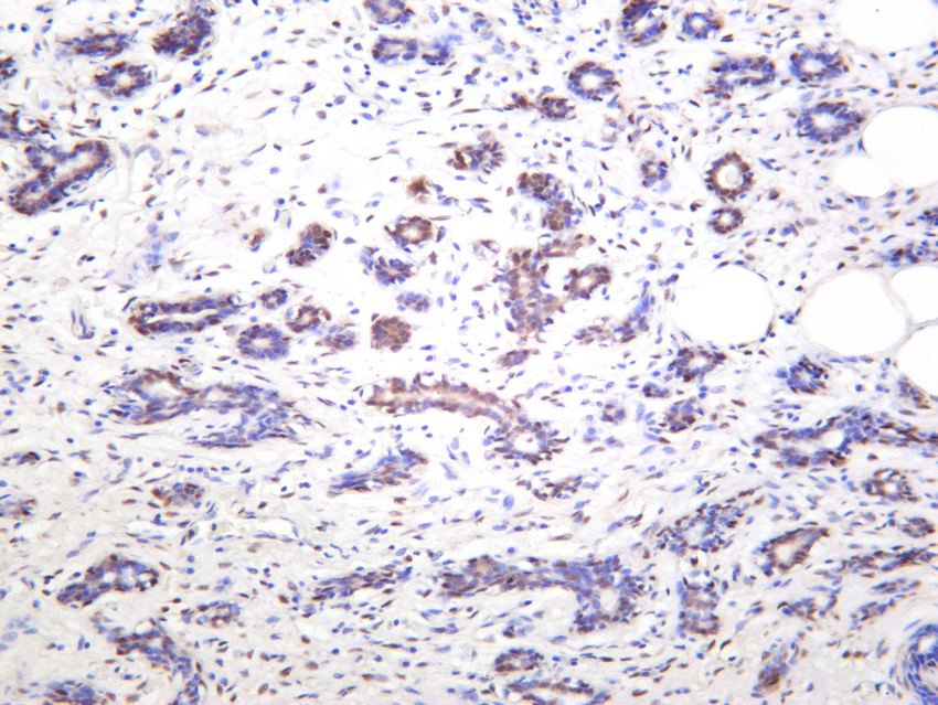
Human breast carcinoma was stained with anti-YAP rabbit antibody.ICC/IF
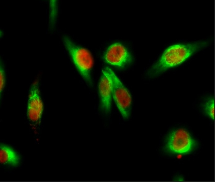
Immunofluorescence analysis of Hela cell. 1,YAP Antibody(red) was diluted at 1:200(4° overnight). GAPDH Monoclonal Antibody(green) was diluted at 1:200(4° overnight). 2, Goat Anti Rabbit Alexa Fluor 594 was diluted at 1:1000(room temperature, 50min). Goat Anti Mouse Alexa Fluor 488 was diluted at 1:1000(room temperature, 50min).| Product Name | YAP Rabbit mAb |
|---|---|
| Antibody Type | Primary Antibodies |
| Clonality | monoclonal |
|---|---|
| Isotype | IgG |
| Host Species | Rabbit |
| Tested Applications | ICC/IFIHCWB |
| WB:1:500-1:2000 IHC:1:100-1:500 ICC/IF:1:200-1:1000 |
|
| Species Reactivity | HumanMouseRat |
| Concentration | 1mg/ml |
| Purification | Protein A |
| Gene Symbol | YAP1 |
|---|---|
| Gene Synonyms | YAP YKI COB1 YAP2 YAP-1 YAP65 |
| Gene Full Name | Yes1 associated transcriptional regulator |
| Gene Summary | This gene encodes a downstream nuclear effector of the Hippo signaling pathway which is involved in development, growth, repair, and homeostasis. This gene is known to play a role in the development and progression of multiple cancers as a transcriptional regulator of this signaling pathway and may function as a potential target for cancer treatment. Alternative splicing results in multiple transcript variants encoding different isoforms. [provided by RefSeq, Aug 2013] |
| Molecular Weight(MW) | 55-75kDa |
| Cellular Localization | Cytoplasm, Nucleus. |
WB
Various whole cell lysates were separated by 4-20% SDS-PAGE, and the membrane was blotted with anti-YAP antibody. The HRP-conjugated Goat anti-Rabbit IgG(H + L) antibody was used to detect the antibody. Lane 1: MCF7, Lane 2: 4T1, Lane 3: C6.
IHC
Mouse thyroid was stained with anti-YAP rabbit antibody.
IHC
Rat thyroid was stained with anti-YAP rabbit antibody.
IHC
Human breast carcinoma was stained with anti-YAP rabbit antibody.
ICC/IF
Immunofluorescence analysis of Hela cell. 1,YAP Antibody(red) was diluted at 1:200(4° overnight). GAPDH Monoclonal Antibody(green) was diluted at 1:200(4° overnight). 2, Goat Anti Rabbit Alexa Fluor 594 was diluted at 1:1000(room temperature, 50min). Goat Anti Mouse Alexa Fluor 488 was diluted at 1:1000(room temperature, 50min).| Application Notes | WB:1:500-1:2000 IHC:1:100-1:500 ICC/IF:1:200-1:1000 |
|---|
| Form | Liquid |
|---|---|
| Storage Instructions | -15°C to -25°C/1 year(Do not lower than -25°C) |
| Storage Buffer | PBS, 50% glycerol, 0.05% Proclin 300, 0.05%BSA |


 Data sheet for OM644255