
WB
Western blot analysis of Phospho-NF-kB p65 (S536) on different lysates with Rabbit anti-Phospho-NF-kB p65 (S536) antibody at 1/5,000 dilution. Lane 1: HeLa cell lysate (20 µg/Lane), Lane 2: HeLa treated with 100nM Calyculin A and 20ng/mL TNF-α for 10 minutes cell lysate (20 µg/Lane), Lane 3: NIH/3T3 cell lysate (20 µg/Lane), Lane 4: NIH/3T3 treated with 100nM Calyculin A and 20ng/mL TNF-α for 10 minutes cell lysate (20 µg/Lane), Exposure time: 59 seconds; 4-20% SDS-PAGE gel. Proteins were transferred to a PVDF membrane and blocked with 5% NFDM/TBST for 1 hour at room temperature. The primary antibody at 1/5,000 dilution was used in 5% NFDM/TBST at 4℃ overnight. Goat Anti-Rabbit IgG - HRP Secondary Antibody at 1/50,000 dilution was used for 1 hour at room temperature.IF
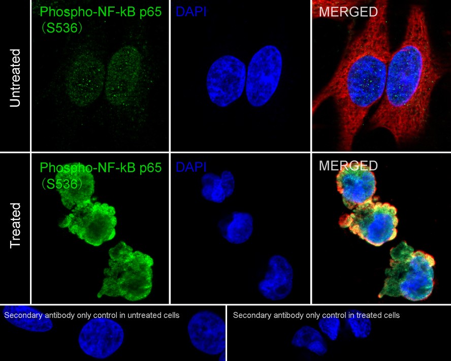
Immunocytochemistry analysis of HeLa cells untreated / treated with 100nM Calyculin A and 20ng/mL TNF-α for 15 minutes labeling Phospho-NF-kB p65 (S536) with Rabbit anti-Phospho-NF-kB p65 (S536) antibody at 1/500 dilution. Cells were fixed in 4% paraformaldehyde for 15 minutes at room temperature, permeabilized with 0.1% Triton X-100 in PBS for 15 minutes at room temperature, then blocked with 1% BSA in 10% negative goat serum for 1 hour at room temperature. Cells were then incubated with Rabbit anti-Phospho-NF-kB p65 (S536) antibody at 1/500 dilution in 1% BSA in PBST overnight at 4 ℃. Goat Anti-Rabbit IgG H&L (488) was used as the secondary antibody at 1/1,000 dilution. PBS instead of the primary antibody was used as the secondary antibody only control. Nuclear DNA was labelled in blue with DAPI. Beta tubulin (red) was stained at 1/100 dilution overnight at +4℃. Goat Anti-Mouse IgG H&L (594) was used as the secondary antibody at 1/1,000 dilution.| Product Name | Phospho-NF-kB p65 (S536) Recombinant Rabbit Monoclonal Antibody |
|---|---|
| Antibody Type | Primary Antibodies |
| Immunogen | Synthetic phospho-peptide corresponding to residues surrounding Ser536 of Human NF-kB p65. |
| Clonality | monoclonal |
|---|---|
| Isotype | IgG |
| Host Species | Rabbit |
| Tested Applications | ICC/IFWB |
| WB:1:5000 ICC/IF:1:500 |
|
| Species Reactivity | HumanMouse |
| Concentration | 1mg/ml |
| Purification | Protein A |
| Gene Symbol | RELA |
|---|---|
| Gene Synonyms | p65 CMCU NFKB3 AIF3BL3 |
| Gene Full Name | RELA proto-oncogene, NF-kB subunit |
| Gene Summary | NF-kappa-B is a ubiquitous transcription factor involved in several biological processes. It is held in the cytoplasm in an inactive state by specific inhibitors. Upon degradation of the inhibitor, NF-kappa-B moves to the nucleus and activates transcription of specific genes. NF-kappa-B is composed of NFKB1 or NFKB2 bound to either REL, RELA, or RELB. The most abundant form of NF-kappa-B is NFKB1 complexed with the product of this gene, RELA. Four transcript variants encoding different isoforms have been found for this gene. [provided by RefSeq, Sep 2011] |
| Molecular Weight(MW) | 60kDa(Observed band size:65kDa) |
| Cellular Localization | Nucleus, Cytoplasm. |
WB
Western blot analysis of Phospho-NF-kB p65 (S536) on different lysates with Rabbit anti-Phospho-NF-kB p65 (S536) antibody at 1/5,000 dilution. Lane 1: HeLa cell lysate (20 µg/Lane), Lane 2: HeLa treated with 100nM Calyculin A and 20ng/mL TNF-α for 10 minutes cell lysate (20 µg/Lane), Lane 3: NIH/3T3 cell lysate (20 µg/Lane), Lane 4: NIH/3T3 treated with 100nM Calyculin A and 20ng/mL TNF-α for 10 minutes cell lysate (20 µg/Lane), Exposure time: 59 seconds; 4-20% SDS-PAGE gel. Proteins were transferred to a PVDF membrane and blocked with 5% NFDM/TBST for 1 hour at room temperature. The primary antibody at 1/5,000 dilution was used in 5% NFDM/TBST at 4℃ overnight. Goat Anti-Rabbit IgG - HRP Secondary Antibody at 1/50,000 dilution was used for 1 hour at room temperature.
IF
Immunocytochemistry analysis of HeLa cells untreated / treated with 100nM Calyculin A and 20ng/mL TNF-α for 15 minutes labeling Phospho-NF-kB p65 (S536) with Rabbit anti-Phospho-NF-kB p65 (S536) antibody at 1/500 dilution. Cells were fixed in 4% paraformaldehyde for 15 minutes at room temperature, permeabilized with 0.1% Triton X-100 in PBS for 15 minutes at room temperature, then blocked with 1% BSA in 10% negative goat serum for 1 hour at room temperature. Cells were then incubated with Rabbit anti-Phospho-NF-kB p65 (S536) antibody at 1/500 dilution in 1% BSA in PBST overnight at 4 ℃. Goat Anti-Rabbit IgG H&L (488) was used as the secondary antibody at 1/1,000 dilution. PBS instead of the primary antibody was used as the secondary antibody only control. Nuclear DNA was labelled in blue with DAPI. Beta tubulin (red) was stained at 1/100 dilution overnight at +4℃. Goat Anti-Mouse IgG H&L (594) was used as the secondary antibody at 1/1,000 dilution.| Application Notes | WB:1:5000 ICC/IF:1:500 |
|---|
| Form | Liquid |
|---|---|
| Storage Instructions | Store at +4℃ after thawing. Aliquot store at -20℃. Avoid repeated freeze / thaw cycles. |
| Storage Buffer | 1*TBS (pH7.4), 0.05% BSA, 40% Glycerol. Preservative: 0.05% Sodium Azide. |


 Data sheet for OM644119