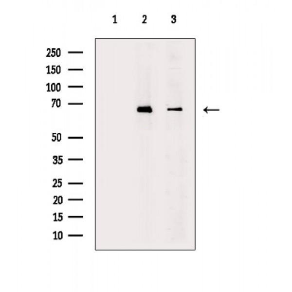
WB
Western blot analysis of extracts from various samples, using Phospho-NF-kB p65 (Ser536) Antibody. Lane 1: HepG2 cells(serum starvation treatment), blocked with antigen-specific peptides, Lane 2: HepG2 cells(serum starvation treatment), Lane 3: RAW264.7 cells(serum starvation treatment).IHC
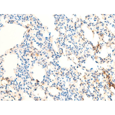
Phospho-NF-kB p65 (Ser536) Antibody at 1/100 staining rat lung tissue sections by IHC-P. The tissue was formaldehyde fixed and a heat mediated antigen retrieval step in citrate buffer was performed. The tissue was then blocked and incubated with the antibody for 1.5 hours at 22°C. An HRP conjugated goat anti-rabbit antibody was used as the secondary antibody.ICC/IF
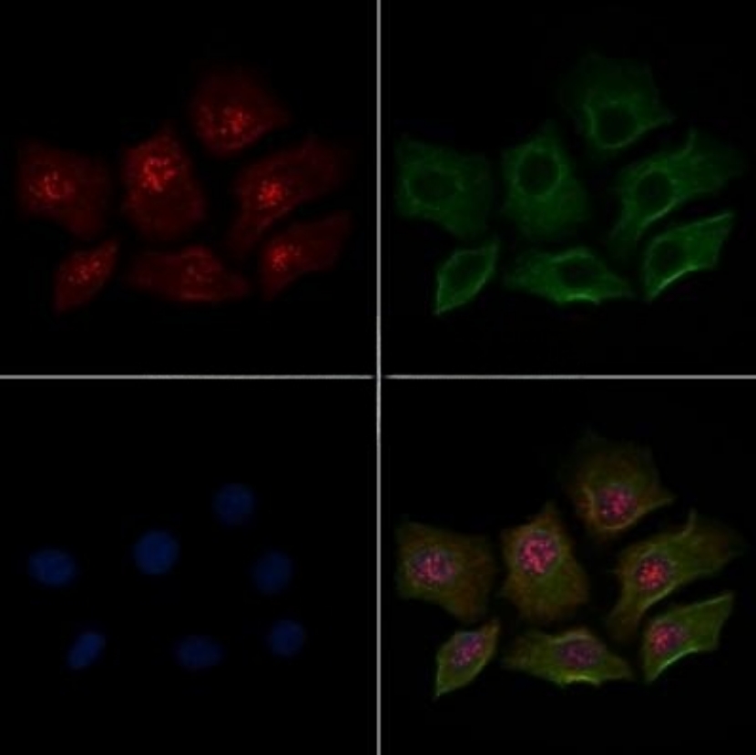
Phospho-NF-kB p65 (Ser536) Antibody staining HepG2 cells(30min of 4uM Forskolin treatment) by ICC/IF. The samples were fixed with PFA and permeabilized in 0.1% Triton X-100,then blocked in 10% serum for 45 minutes at 25°C. Samples were then incubated with primary Ab and mouse anti-beta tubulin Ab for 1 hour at 37°C. An AlexaFluor594 conjugated goat anti-rabbit IgG(H+L) Ab(Red) and an AlexaFluor488 conjugated goat anti-mouse IgG(H+L) Ab(Green) were used as the secondary antibody. The nuclear counter stain is DAPI(blue).| Product Name | Rabbit polyclonal antibody to Phospho-NF-kB p65 (Ser536) |
|---|---|
| Antibody Type | Primary Antibodies |
| Immunogen | A synthesized peptide derived from human RELA(Accession Q04206), corresponding to amino acid residues around phosphorylated Ser536. |
| Clonality | polyclonal |
|---|---|
| Isotype | IgG |
| Host Species | Rabbit |
| Tested Applications | ICC/IFIHCWB |
| WB:1:500-1:2000 IHC:1:50-1:500 ICC/IF:1:200 |
|
| Species Reactivity | HumanMonkeyRat |
| Concentration | 1mg/ml |
| Purification | Affinity purified |
| Gene Symbol | RELA |
|---|---|
| Gene Synonyms | p65 CMCU NFKB3 AIF3BL3 |
| Gene Full Name | RELA proto-oncogene, NF-kB subunit |
| Gene Summary | NF-kappa-B is a ubiquitous transcription factor involved in several biological processes. It is held in the cytoplasm in an inactive state by specific inhibitors. Upon degradation of the inhibitor, NF-kappa-B moves to the nucleus and activates transcription of specific genes. NF-kappa-B is composed of NFKB1 or NFKB2 bound to either REL, RELA, or RELB. The most abundant form of NF-kappa-B is NFKB1 complexed with the product of this gene, RELA. Four transcript variants encoding different isoforms have been found for this gene. [provided by RefSeq, Sep 2011] |
| Molecular Weight(MW) | 65kDa; 60kD(Calculated). |
| Cellular Localization | Nucleus, Cytoplasm. |
WB
Western blot analysis of extracts from various samples, using Phospho-NF-kB p65 (Ser536) Antibody. Lane 1: HepG2 cells(serum starvation treatment), blocked with antigen-specific peptides, Lane 2: HepG2 cells(serum starvation treatment), Lane 3: RAW264.7 cells(serum starvation treatment).
IHC
Phospho-NF-kB p65 (Ser536) Antibody at 1/100 staining rat lung tissue sections by IHC-P. The tissue was formaldehyde fixed and a heat mediated antigen retrieval step in citrate buffer was performed. The tissue was then blocked and incubated with the antibody for 1.5 hours at 22°C. An HRP conjugated goat anti-rabbit antibody was used as the secondary antibody.
ICC/IF
Phospho-NF-kB p65 (Ser536) Antibody staining HepG2 cells(30min of 4uM Forskolin treatment) by ICC/IF. The samples were fixed with PFA and permeabilized in 0.1% Triton X-100,then blocked in 10% serum for 45 minutes at 25°C. Samples were then incubated with primary Ab and mouse anti-beta tubulin Ab for 1 hour at 37°C. An AlexaFluor594 conjugated goat anti-rabbit IgG(H+L) Ab(Red) and an AlexaFluor488 conjugated goat anti-mouse IgG(H+L) Ab(Green) were used as the secondary antibody. The nuclear counter stain is DAPI(blue).| Application Notes | WB:1:500-1:2000 IHC:1:50-1:500 ICC/IF:1:200 |
|---|
| Form | Liquid |
|---|---|
| Storage Instructions | Store at -20 °C. Stable for 12 months from date of receipt. |
| Storage Buffer | Rabbit IgG in phosphate buffered saline , pH 7.4, 150mM NaCl, 0.02% sodium azide and 50% glycerol. |


 Data sheet for OM644121