WB
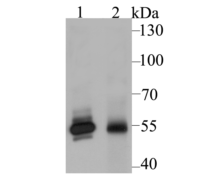
Western blot analysis of GLUR on different lysates using anti-GLUR antibody at 1/2,000 dilution. Positive control: Lane 1: A549, Lane 2: Rat liver tissue.IHC
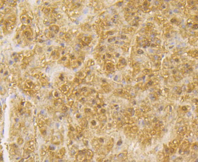
Immunohistochemical analysis of paraffin-embedded human liver tissue using anti-GLUR antibody. Counter stained with hematoxylin.IHC
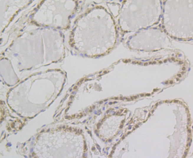
Immunohistochemical analysis of paraffin-embedded human thyroid gland tissue using anti-GLUR antibody. Counter stained with hematoxylin.ICC/IF
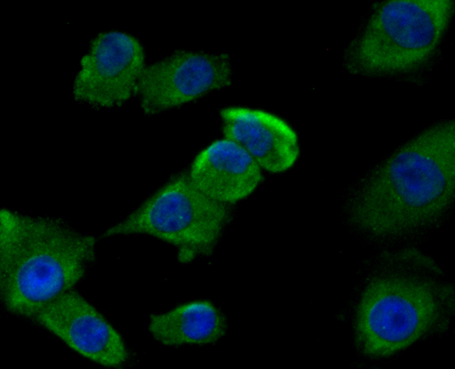
ICC staining GLUR in A549 cells (green). The nuclear counter stain is DAPI (blue). Cells were fixed in paraformaldehyde, permeabilised with 0.25% Triton X100/PBS.FC
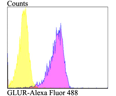
Flow cytometric analysis of A549 cells with GLUR antibody at 1/100 dilution (purple) compared with an unlabelled control (cells without incubation with primary antibody; yellow). Alexa Fluor 488-conjugated goat anti-rabbit IgG was used as the secondary antibody.| Product Name | Glutathione Reductase Rabbit Polyclonal Antibody |
|---|---|
| Antibody Type | Primary Antibodies |
| Immunogen | Recombinant protein within Human GLUR aa 273-446 / 522. |
| Clonality | polyclonal |
|---|---|
| Isotype | IgG |
| Host Species | Rabbit |
| Tested Applications | FCICC/IFIHCWB |
| WB:1:1000-1:2000 IHC:1:50-1:200 ICC/IF:1:50-1:200 FC:1:50-1:100 |
|
| Species Reactivity | HumanRat |
| Concentration | 1mg/ml |
| Purification | Affinity purified |
| Gene Symbol | GSR |
|---|---|
| Gene Synonyms | GR GSRD HEL-75 CNSHA10 HEL-S-122m |
| Gene Full Name | glutathione-disulfide reductase |
| Gene Summary | This gene encodes a member of the class-I pyridine nucleotide-disulfide oxidoreductase family. This enzyme is a homodimeric flavoprotein. It is a central enzyme of cellular antioxidant defense, and reduces oxidized glutathione disulfide (GSSG) to the sulfhydryl form GSH, which is an important cellular antioxidant. Rare mutations in this gene result in hereditary glutathione reductase deficiency. Multiple alternatively spliced transcript variants encoding different isoforms have been found. [provided by RefSeq, Aug 2010] |
| Molecular Weight(MW) | 56 KDa |
| Cellular Localization | Cytoplasm. Mitochondrion. |
WB
Western blot analysis of GLUR on different lysates using anti-GLUR antibody at 1/2,000 dilution. Positive control: Lane 1: A549, Lane 2: Rat liver tissue.
IHC
Immunohistochemical analysis of paraffin-embedded human liver tissue using anti-GLUR antibody. Counter stained with hematoxylin.
IHC
Immunohistochemical analysis of paraffin-embedded human thyroid gland tissue using anti-GLUR antibody. Counter stained with hematoxylin.
ICC/IF
ICC staining GLUR in A549 cells (green). The nuclear counter stain is DAPI (blue). Cells were fixed in paraformaldehyde, permeabilised with 0.25% Triton X100/PBS.
FC
Flow cytometric analysis of A549 cells with GLUR antibody at 1/100 dilution (purple) compared with an unlabelled control (cells without incubation with primary antibody; yellow). Alexa Fluor 488-conjugated goat anti-rabbit IgG was used as the secondary antibody.| Application Notes | WB:1:1000-1:2000 IHC:1:50-1:200 ICC/IF:1:50-1:200 FC:1:50-1:100 |
|---|
| Form | Liquid |
|---|---|
| Storage Instructions | Store at +4℃ after thawing. Aliquot store at -20℃. Avoid repeated freeze / thaw cycles. |
| Storage Buffer | 1*PBS (pH7.4), 0.2% BSA, 50% Glycerol. Preservative: 0.05% Sodium Azide. |


 Data sheet for OM644176