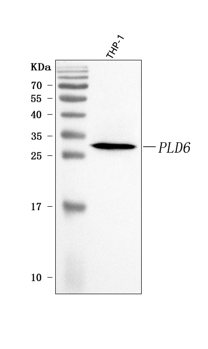
WB
Western blot analysis of anti-PLD6 antibody. The sample well of each lane was loaded with 30 ug of sample under reducing conditions. Lane 1: human THP-1 whole cell lysates. After electrophoresis, proteins were transferred to a membrane. Then the membrane was incubated with rabbit anti-PLD6 antigen affinity purified polyclonal antibody and probed with a goat anti-rabbit IgG-HRP secondary antibody. The signal is developed using ECL Plus Western Blotting Substrate.IHC
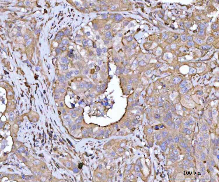
IHC analysis of PLD6 using anti-PLD6 antibody. PLD6 was detected in a paraffin-embedded section of human colon adenocarcinoma tissue. The tissue section was developed using HRP Conjugated Rabbit IgG Super Vision Assay Kit with DAB as the chromogen.IF-P
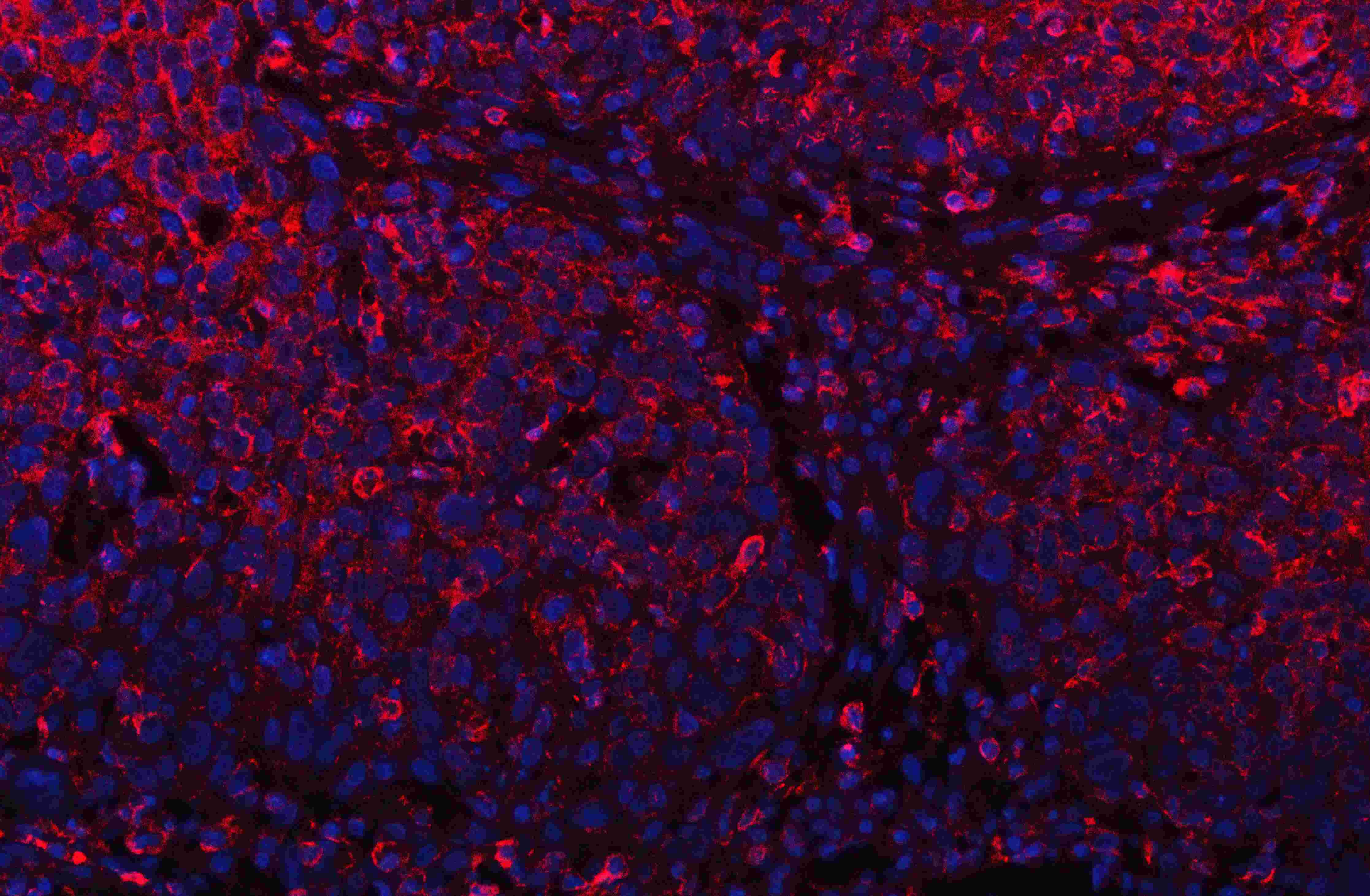
IF analysis of PLD6 using anti-PLD6 antibody. PLD6 was detected in a paraffin-embedded section of human oesophagus squama cancer tissue. Cy3-conjugated Anti-rabbit IgG Secondary Antibody (red)was used as secondary antibody. The section was counterstained with DAPI (Blue).FC
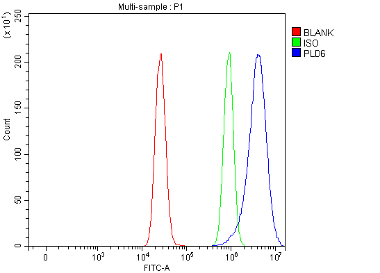
Flow Cytometry analysis of MCF-7 cells using anti-PLD6 antibody. Overlay histogram showing MCF-7 cells stained withanti-PLD6 antibody (Blue line). To facilitate intracellular staining, cells were fixed with 4% paraformaldehyde and permeabilized with permeabilization buffer. The cells were blocked with 10% normal goat serum. And then incubated with rabbit anti-PLD6 Antibody (1:100). DyLight 488 conjugated goat anti-rabbit IgG (1:100) was used as secondary antibody. Isotype control antibody (Green line) was rabbit IgG(1:100) used under the same conditions. Unlabelled sample (Red line) was also used as a control.| Product Name | Rabbit polyclonal antibody to PLD6 |
|---|---|
| Antibody Type | Primary Antibodies |
| Immunogen | E. coli-derived human PLD6 recombinant protein (Position: R39-L238). Human PLD6 shares 87.9% amino acid (aa) sequence identity with mouse PLD6. |
| Clonality | polyclonal |
|---|---|
| Isotype | IgG |
| Host Species | Rabbit |
| Tested Applications | FCIF-PIHCWB |
| WB:1:500-1:2000 IHC:1:50-1:400 IF-P:1:50-1:400 FC:1:50-1:200 |
|
| Species Reactivity | Human |
| Concentration | 0.5mg/ml |
| Purification | Affinity purified |
| Gene Symbol | PLD6 |
|---|---|
| Gene Synonyms | ZUC |
| Gene Full Name | phospholipase D family member 6 |
| Gene Summary | Enables cardiolipin hydrolase activity and protein homodimerization activity. Involved in mitochondrial fusion. Acts upstream of or within positive regulation of mitochondrial fusion. Located in mitochondrial outer membrane. [provided by Alliance of Genome Resources, Mar 2025] |
| Molecular Weight(MW) | 28kDa |
| Cellular Localization | Mitochondrion outer membrane, Golgi apparatus. |
WB
Western blot analysis of anti-PLD6 antibody. The sample well of each lane was loaded with 30 ug of sample under reducing conditions. Lane 1: human THP-1 whole cell lysates. After electrophoresis, proteins were transferred to a membrane. Then the membrane was incubated with rabbit anti-PLD6 antigen affinity purified polyclonal antibody and probed with a goat anti-rabbit IgG-HRP secondary antibody. The signal is developed using ECL Plus Western Blotting Substrate.
IHC
IHC analysis of PLD6 using anti-PLD6 antibody. PLD6 was detected in a paraffin-embedded section of human colon adenocarcinoma tissue. The tissue section was developed using HRP Conjugated Rabbit IgG Super Vision Assay Kit with DAB as the chromogen.
IF-P
IF analysis of PLD6 using anti-PLD6 antibody. PLD6 was detected in a paraffin-embedded section of human oesophagus squama cancer tissue. Cy3-conjugated Anti-rabbit IgG Secondary Antibody (red)was used as secondary antibody. The section was counterstained with DAPI (Blue).
FC
Flow Cytometry analysis of MCF-7 cells using anti-PLD6 antibody. Overlay histogram showing MCF-7 cells stained withanti-PLD6 antibody (Blue line). To facilitate intracellular staining, cells were fixed with 4% paraformaldehyde and permeabilized with permeabilization buffer. The cells were blocked with 10% normal goat serum. And then incubated with rabbit anti-PLD6 Antibody (1:100). DyLight 488 conjugated goat anti-rabbit IgG (1:100) was used as secondary antibody. Isotype control antibody (Green line) was rabbit IgG(1:100) used under the same conditions. Unlabelled sample (Red line) was also used as a control.| Application Notes | WB:1:500-1:2000 IHC:1:50-1:400 IF-P:1:50-1:400 FC:1:50-1:200 |
|---|
| Form | Liquid |
|---|---|
| Storage Instructions | 12 months from date of receipt, -20℃ as supplied. 6 months 2 to 8℃ after reconstitution. Avoid repeated freezing and thawing. |
| Storage Buffer | 500ug/ml antibody with PBS, 0.02% NaN3, 1 mg/ml BSA and 50% glycerol. |


 Data sheet for OM644177