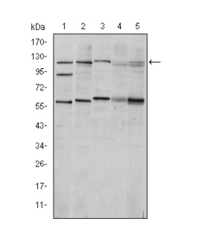

WB
Western blot analysis using HIF1A mouse mAb against Cos7 (1), Hela (2), Jurkat (3), RAJI (4) and NIH/3T3 (5) cell lysate.IHC
Immunohistochemical analysis of paraffin-embedded stomach cancer tissues (left) and brain tumor tissues (right) using HIF1A mouse mAb with DAB staining.ICC/IF
Immunofluorescence analysis of Hela cells using HIF1A mouse mAb (green). Blue: DRAQ5 fluorescent DNA dye. Red: Actin filaments have been labeled with Alexa Fluor-555 phalloidin.| Product Name | Mouse Monoclonal to HIF1A |
|---|---|
| Antibody Type | Primary Antibodies |
| Immunogen | Purified recombinant fragment of human HIF1A expressed in E. Coli. |
| Clonality | monoclonal |
|---|---|
| Isotype | IgG1 |
| Host Species | Mouse |
| Tested Applications | ICC/IFIHCWB |
| WB:1:500-1:2000 IHC:1:200-1:1000 ICC/IF:1:200-1:1000 |
|
| Species Reactivity | HumanMonkeyMouse |
| Concentration | 1mg/ml |
| Purification | Affinity purified |
| Gene Symbol | HIF1A |
|---|---|
| Gene Synonyms | HIF1 MOP1 PASD8 HIF-1A bHLHe78 HIF-1alpha HIF1-ALPHA HIF-1-alpha |
| Gene Full Name | hypoxia inducible factor 1 subunit alpha |
| Gene Summary | This gene encodes the alpha subunit of transcription factor hypoxia-inducible factor-1 (HIF-1), which is a heterodimer composed of an alpha and a beta subunit. HIF-1 functions as a master regulator of cellular and systemic homeostatic response to hypoxia by activating transcription of many genes, including those involved in energy metabolism, angiogenesis, apoptosis, and other genes whose protein products increase oxygen delivery or facilitate metabolic adaptation to hypoxia. HIF-1 thus plays an essential role in embryonic vascularization, tumor angiogenesis and pathophysiology of ischemic disease. Alternatively spliced transcript variants encoding different isoforms have been identified for this gene. [provided by RefSeq, Jul 2011] |
| Molecular Weight(MW) | 120kDa |
| Cellular Localization | Cytoplasm, Nucleus |
WB
Western blot analysis using HIF1A mouse mAb against Cos7 (1), Hela (2), Jurkat (3), RAJI (4) and NIH/3T3 (5) cell lysate.
IHC
Immunohistochemical analysis of paraffin-embedded stomach cancer tissues (left) and brain tumor tissues (right) using HIF1A mouse mAb with DAB staining.
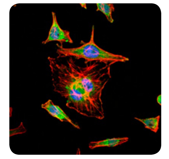
ICC/IF
Immunofluorescence analysis of Hela cells using HIF1A mouse mAb (green). Blue: DRAQ5 fluorescent DNA dye. Red: Actin filaments have been labeled with Alexa Fluor-555 phalloidin.| Application Notes | WB:1:500-1:2000 IHC:1:200-1:1000 ICC/IF:1:200-1:1000 |
|---|
| Form | Liquid |
|---|---|
| Storage Instructions | 4°C; -20°C for long term storage |
| Storage Buffer | Ascitic fluid containing 0.03% sodium azide. |


 Data sheet for OM650009