
Application
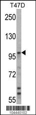
Western blot analysis of UBE3A Antibody (C-term) in T47D cell line lysates (35ug/lane).UBE3A(arrow) was detected using the purified Pab.Application
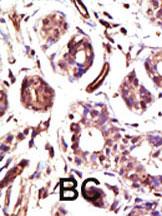
Formalin-fixed and paraffin-embedded human cancer tissue reacted with the primary antibody, which was peroxidase-conjugated to the secondary antibody, followed by DAB staining. This data demonstrates the use of this antibody for immunohistochemistry; clinical relevance has not been evaluated. BC = breast carcinoma; HC = hepatocarcinoma.Application
Confocal immunofluorescent analysis of UBE3A Antibody (C-term)(Cat#AP2154b) with Hela cell followed by Alexa Fluor 488-conjugated goat anti-rabbit lgG (green). Actin filaments have been labeled with Alexa Fluor 555 phalloidin (red).DAPI was used to stain the cell nuclear (blue).Application
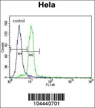
UBE3A Antibody (C-term) flow cytometric analysis of Hela cells (right histogram) compared to a negative control cell (left histogram).FITC-conjugated goat-anti-rabbit secondary antibodies were used for the analysis.| Product Name | UBE3A Antibody (C-term) |
|---|---|
| Antibody Type | Primary Antibodies |
| Antigen Alias | UBE3A; E6AP; EPVE6AP; HPVE6A; Ubiquitin-protein ligase E3A; E6AP ubiquitin-protein ligase; Human papillomavirus E6-associated protein; Oncogenic protein-associated protein E6-AP; Renal carcinoma antigen NY-REN-54 |
| Clonality | Polyclonal |
|---|---|
| Isotype | Ig |
| Host Species | Rabbit |
| Tested Applications | WBIHCIFFC |
| WB:1:100~500 IHC |
|
| Species Reactivity | Human |
| Concentration | 1mg/ml |
| Gene Synonyms | E6AP EPVE6AP HPVE6A |
|---|---|
| Alternative Names | UBE3A E6AP EPVE6AP HPVE6A Ubiquitin-protein ligase E3A E6AP ubiquitin-protein ligase Human papillomavirus E6-associated protein Oncogenic protein-associated protein E6-AP Renal carcinoma antigen NY-REN-54 |
| Molecular Weight(MW) | 100688 Da |
| Function | E3 ubiquitin-protein ligase which accepts ubiquitin from an E2 ubiquitin-conjugating enzyme in the form of a thioester and transfers it to its substrates. Several substrates have been identified including the RAD23A and RAD23B, MCM7 (which is involved in DNA replication), annexin A1, the PML tumor suppressor, and the cell cycle regulator CDKN1B. Catalyzes the high-risk human papilloma virus E6-mediated ubiquitination of p53/TP53, contributing to the neoplastic progression of cells infected by these viruses. Additionally, may function as a cellular quality control ubiquitin ligase by helping the degradation of the cytoplasmic misfolded proteins. Finally, UBE3A also promotes its own degradation in vivo |
| Tissue Specificity | This UBE3A antibody is generated from rabbits immunized with a KLH conjugated synthetic peptide between 843~873 amino acids from the C-terminal region of human UBE3A. |
| Cellular Localization | Nucleus (Probable). |
| Entrez Gene | 7337 |
|---|
Application
Western blot analysis of UBE3A Antibody (C-term) in T47D cell line lysates (35ug/lane).UBE3A(arrow) was detected using the purified Pab.
Application
Formalin-fixed and paraffin-embedded human cancer tissue reacted with the primary antibody, which was peroxidase-conjugated to the secondary antibody, followed by DAB staining. This data demonstrates the use of this antibody for immunohistochemistry; clinical relevance has not been evaluated. BC = breast carcinoma; HC = hepatocarcinoma.
Application
Confocal immunofluorescent analysis of UBE3A Antibody (C-term)(Cat#AP2154b) with Hela cell followed by Alexa Fluor 488-conjugated goat anti-rabbit lgG (green). Actin filaments have been labeled with Alexa Fluor 555 phalloidin (red).DAPI was used to stain the cell nuclear (blue).
Application
UBE3A Antibody (C-term) flow cytometric analysis of Hela cells (right histogram) compared to a negative control cell (left histogram).FITC-conjugated goat-anti-rabbit secondary antibodies were used for the analysis.| Application Notes | WB:1:100~500 IHC |
|---|
| Form | Liquid |
|---|---|
| Storage Instructions | For short-term storage, store at 4° C. For long-term storage, aliquot and store at -20ºC or below. Avoid multiple freeze-thaw cycles. |
| Storage Buffer | Purified polyclonal antibody supplied in PBS with 0.09% (W/V) sodium azide. This antibody is purified through a protein G column, eluted with high and low pH buffers and neutralized immediately, followed by dialysis against PBS. |


 Data sheet for OM224106