Application
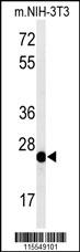
Western blot analysis of CAV1 Antibody (Center) in mouse NIH-3T3 cell line lysates (35ug/lane). CAV1 (arrow) was detected using the purified Pab.Application
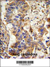
CAV1 Antibody (Center) IHC analysis in formalin fixed and paraffin embedded lung carcinoma followed by peroxidase conjugation of the secondary antibody and DAB staining. This data demonstrates the use of the CAV1 Antibody (Center) for immunohistochemistry. Clinical relevance has not been evaluated.Application
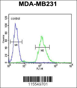
CAV1 Antibody (Center) flow cytometric analysis of MDA-MB231 cells (right histogram) compared to a negative control cell (left histogram).FITC-conjugated goat-anti-rabbit secondary antibodies were used for the analysis.Application
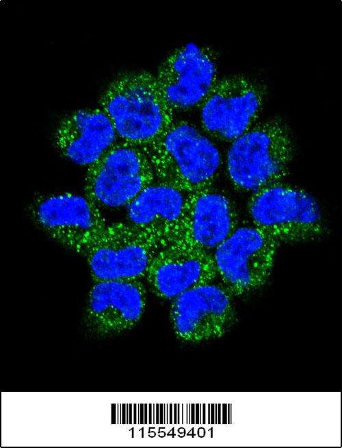
Confocal immunofluorescent analysis of CAV1 Antibody (Center)(Cat#AP7767c) with NCI-H460 cell followed by Alexa Fluor 488-conjugated goat anti-rabbit lgG (green).DAPI was used to stain the cell nuclear (blue).| Product Name | CAV1 Antibody (Center) |
|---|---|
| Antibody Type | Primary Antibodies |
| Antigen Alias | CAV1; CAV; Caveolin-1 |
| Clonality | Polyclonal |
|---|---|
| Isotype | Ig |
| Host Species | Rabbit |
| Tested Applications | WBIHCFCIF |
| WB:1:100~500 IHC |
|
| Species Reactivity | Human |
| Concentration | 1mg/ml |
| Gene Synonyms | CAV |
|---|---|
| Alternative Names | CAV1 CAV Caveolin-1 |
| Molecular Weight(MW) | 20472 Da |
| Function | May act as a scaffolding protein within caveolar membranes. Interacts directly with G-protein alpha subunits and can functionally regulate their activity (By similarity). Involved in the costimulatory signal essential for T-cell receptor (TCR)- mediated T-cell activation. Its binding to DPP4 induces T-cell proliferation and NF-kappa-B activation in a T-cell receptor/CD3- dependent manner. Recruits CTNNB1 to caveolar membranes and may regulate CTNNB1-mediated signaling through the Wnt pathway |
| Tissue Specificity | Expressed in muscle and lung, less so in liver, brain and kidney |
| Cellular Localization | Golgi apparatus membrane; Peripheral membrane protein. Cell membrane; Peripheral membrane protein Membrane, caveola; Peripheral membrane protein. Membrane raft Note=Colocalized with DPP4 in membrane rafts. Potential hairpin- like structure in the membrane. Membrane protein of caveolae |
| Entrez Gene | 857 |
|---|
Application
Western blot analysis of CAV1 Antibody (Center) in mouse NIH-3T3 cell line lysates (35ug/lane). CAV1 (arrow) was detected using the purified Pab.
Application
CAV1 Antibody (Center) IHC analysis in formalin fixed and paraffin embedded lung carcinoma followed by peroxidase conjugation of the secondary antibody and DAB staining. This data demonstrates the use of the CAV1 Antibody (Center) for immunohistochemistry. Clinical relevance has not been evaluated.
Application
CAV1 Antibody (Center) flow cytometric analysis of MDA-MB231 cells (right histogram) compared to a negative control cell (left histogram).FITC-conjugated goat-anti-rabbit secondary antibodies were used for the analysis.
Application
Confocal immunofluorescent analysis of CAV1 Antibody (Center)(Cat#AP7767c) with NCI-H460 cell followed by Alexa Fluor 488-conjugated goat anti-rabbit lgG (green).DAPI was used to stain the cell nuclear (blue).| Application Notes | WB:1:100~500 IHC |
|---|
| Form | Liquid |
|---|---|
| Storage Instructions | For short-term storage, store at 4° C. For long-term storage, aliquot and store at -20ºC or below. Avoid multiple freeze-thaw cycles. |
| Storage Buffer | Purified polyclonal antibody supplied in PBS with 0.09% (W/V) sodium azide. This antibody is prepared by Saturated Ammonium Sulfate (SAS) precipitation followed by dialysis against PBS. |


 Data sheet for OM228786