Application
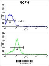
Flow cytometric analysis of MCF-7 cells (bottom histogram) compared to a negative control cell (top histogram).FITC-conjugated goat-anti-rabbit secondary antibodies were used for the analysis.Application
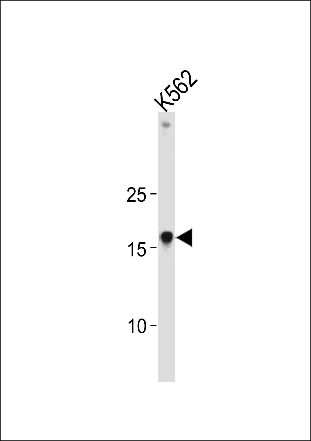
Western blot analysis in K562 cell line lysates (35ug/lane).Application
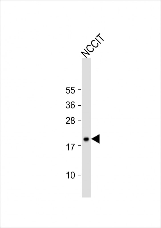
Western Blot at 1:1000 dilution + NCCIT whole cell lysate Lysates/proteins at 20 ug per lane.Application
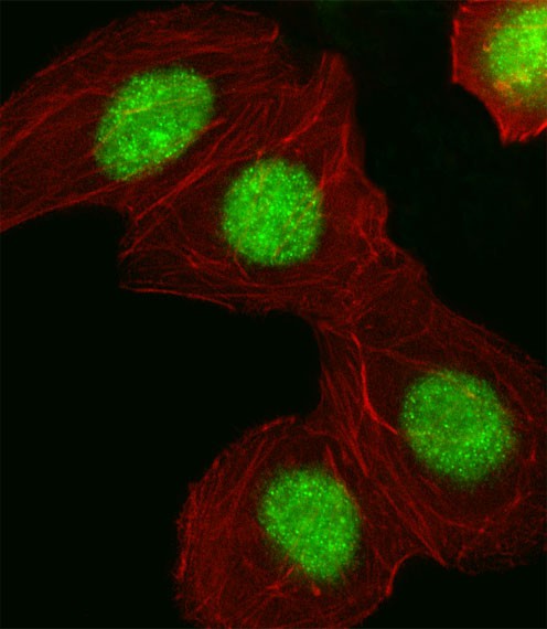
Fluorescent image of A549 cell stained with HMGA1 Antibody .A549 cells were fixed with 4% PFA (20 min), permeabilized with Triton X-100 (0.1%, 10 min), then incubated with HMGA1 primary antibody (1:25). For secondary antibody, Alexa Fluor 488 conjugated donkey anti-rabbit antibody (green) was used (1:400).Cytoplasmic actin was counterstained with Alexa Fluor 555 (red) conjugated Phalloidin (7units| Product Name | HMGA1 Antibody |
|---|---|
| Antibody Type | Primary Antibodies |
| Antigen Alias | High mobility group protein HMG-I/HMG-Y, HMG-I(Y), High mobility group AT-hook protein 1, High mobility group protein A1, High mobility group protein R, HMGA1, HMGIY |
| Product description | HMGA1 encodes a non-histone protein involved in many cellular processes, including regulation of inducible gene transcription, integration of retroviruses into chromosomes, and the metastatic progression of cancer cells. The encoded protein preferentially binds to the minor groove of A+T-rich regions in double-stranded DNA. It has little secondary structure in solution but assumes distinct conformations when bound to substrates such as DNA or other proteins. The encoded protein is frequently acetylated and is found in the nucleus.1) Mu,G., et.al., Hum. Pathol. 41 (4), 493-502 (2010) |
| Immunogen | This HMGA1 antibody is generated from rabbits immunized with a KLH conjugated synthetic peptide between 64-93 amino acids from the C-terminal region of human HMGA1. |
| Clonality | Polyclonal |
|---|---|
| Isotype | Ig |
| Host Species | Rabbit |
| Tested Applications | FACSIFWB |
| For WB starting dilution is: 1:1000 For IF starting dilution is: 1:10~50 For FACS starting dilution is: 1:10~50: |
|
| Species Reactivity | Human |
| Concentration | 1mg/ml |
| Gene Symbol | HMGA1 |
|---|---|
| Alternative Names | High mobility group protein HMG-I/HMG-Y HMG-I(Y) High mobility group AT-hook protein 1 High mobility group protein A1 High mobility group protein R HMGA1 HMGIY |
| Molecular Weight(MW) | 12 kDa |
| Sequence Similarities | Predicted species reactivity based on immunogen sequence: Hamster |
Application
Flow cytometric analysis of MCF-7 cells (bottom histogram) compared to a negative control cell (top histogram).FITC-conjugated goat-anti-rabbit secondary antibodies were used for the analysis.
Application
Western blot analysis in K562 cell line lysates (35ug/lane).
Application
Western Blot at 1:1000 dilution + NCCIT whole cell lysate Lysates/proteins at 20 ug per lane.
Application
Fluorescent image of A549 cell stained with HMGA1 Antibody .A549 cells were fixed with 4% PFA (20 min), permeabilized with Triton X-100 (0.1%, 10 min), then incubated with HMGA1 primary antibody (1:25). For secondary antibody, Alexa Fluor 488 conjugated donkey anti-rabbit antibody (green) was used (1:400).Cytoplasmic actin was counterstained with Alexa Fluor 555 (red) conjugated Phalloidin (7units| Application Notes | For WB starting dilution is: 1:1000 For IF starting dilution is: 1:10~50 For FACS starting dilution is: 1:10~50: |
|---|
| Form | Liquid |
|---|---|
| Storage Instructions | Store at 4˚C for three months and -20˚C, stable for up to one year. As with all antibodies care should be taken to avoid repeated freeze thaw cycles. Antibodies should not be exposed to prolonged high temperatures. |
| Storage Buffer | Supplied in PBS with 0.09% (W/V) sodium azide. |


 Data sheet for OM281076