WB
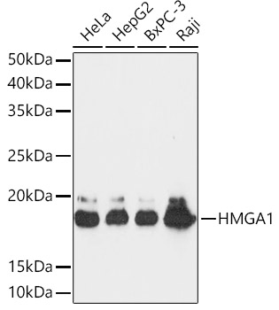
Western blot analysis of various lysates using HMGA1 Rabbit mAb at 1:1000 dilution. Secondary antibody: HRP-conjugated Goat anti-Rabbit IgG (H+L) at 1:10000 dilution. Lysates/proteins: 25μg per lane. Blocking buffer: 3% nonfat dry milk in TBST. Detection: ECL Basic Kit. Exposure time: 30s.IHC
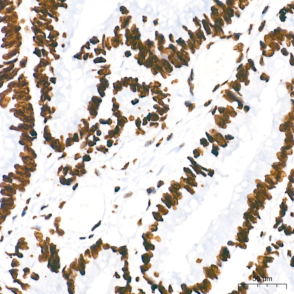
Immunohistochemistry analysis of paraffin embedded Human colon carcinoma tissue using HMGA1 Rabbit mAb at a dilution of 1:5000 (40x lens). High pressure antigen retrieval performed with 0.01M Tris EDTA Buffer (pH 9.0) prior to IHC staining.ICC/IF
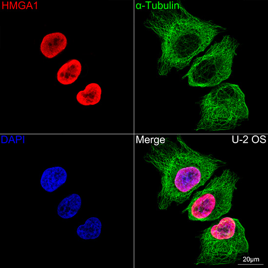
Confocal imaging of U-2OS cells using HMGA1 Rabbit mAb (dilution 1:100)(Red). The cells were counterstained with α-Tubulin Mouse mAb (dilution 1:400) (Green). DAPI was used for nuclear staining (blue). objective: 100x.| Product Name | HMGA1 Rabbit mAb |
|---|---|
| Antibody Type | Primary Antibodies |
| Immunogen | A synthetic peptide corresponding to a sequence within amino acids 1-107 of human HMGA1 (P17096). |
| Clonality | Monoclonal |
|---|---|
| Isotype | IgG |
| Host Species | Rabbit |
| Tested Applications | ICC/IFIHCWB |
| WB:1:1000-1:4000 IHC:1:2000-1:8000 ICC:1:100-1:1000 |
|
| Species Reactivity | HumanMouseRat |
| Concentration | 1mg/ml |
| Purification | Affinity purified |
| Gene Symbol | HMGA1 |
|---|---|
| Gene Synonyms | HMG-R HMGIY HMGA1A |
| Gene Full Name | high mobility group AT-hook 1 |
| Gene Summary | This gene encodes a chromatin-associated protein involved in the regulation of gene transcription, integration of retroviruses into chromosomes, and the metastatic progression of cancer cells. The encoded protein preferentially binds to the minor groove of AT-rich regions in double-stranded DNA. Multiple transcript variants encoding different isoforms have been found for this gene. Pseudogenes of this gene have been identified on multiple chromosomes. [provided by RefSeq, Jan 2016] |
| Molecular Weight(MW) | 12kDa(Observed MW 18kDa) |
| Cellular Localization | Chromosome,Nucleus. |
WB
Western blot analysis of various lysates using HMGA1 Rabbit mAb at 1:1000 dilution. Secondary antibody: HRP-conjugated Goat anti-Rabbit IgG (H+L) at 1:10000 dilution. Lysates/proteins: 25μg per lane. Blocking buffer: 3% nonfat dry milk in TBST. Detection: ECL Basic Kit. Exposure time: 30s.
IHC
Immunohistochemistry analysis of paraffin embedded Human colon carcinoma tissue using HMGA1 Rabbit mAb at a dilution of 1:5000 (40x lens). High pressure antigen retrieval performed with 0.01M Tris EDTA Buffer (pH 9.0) prior to IHC staining.
ICC/IF
Confocal imaging of U-2OS cells using HMGA1 Rabbit mAb (dilution 1:100)(Red). The cells were counterstained with α-Tubulin Mouse mAb (dilution 1:400) (Green). DAPI was used for nuclear staining (blue). objective: 100x.| Application Notes | WB:1:1000-1:4000 IHC:1:2000-1:8000 ICC:1:100-1:1000 |
|---|
| Form | Liquid |
|---|---|
| Storage Instructions | Store at -20℃. Avoid freeze / thaw cycles. |
| Storage Buffer | Buffer: PBS with 0.02% sodium azide,0.05% BSA,50% glycerol,pH7.3. |


 Data sheet for OM643538