Application
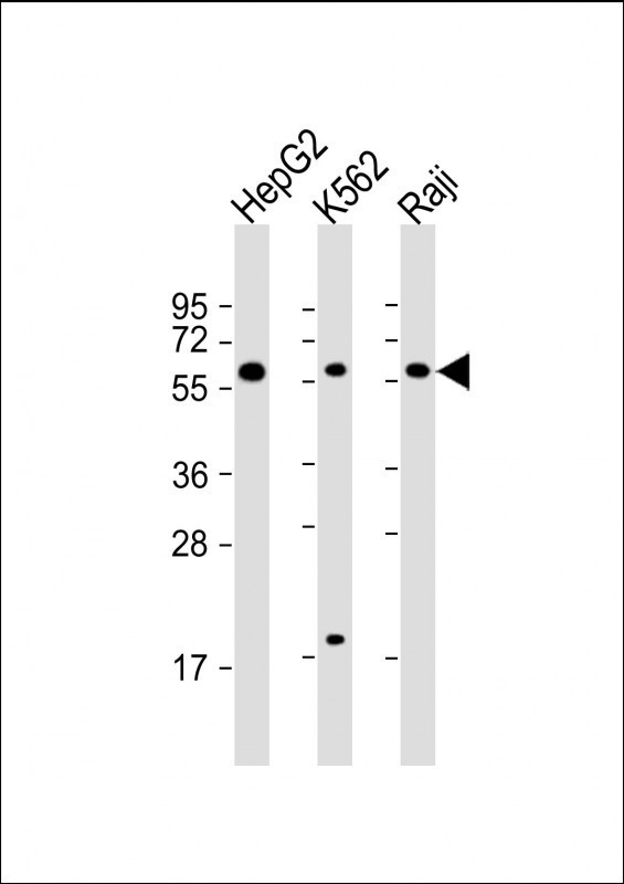
Western Blot at 1:2000 dilution Lane 1: HepG2 whole cell lysate Lane 2: K562 whole cell lysate Lane 3: Raji whole cell lysate Lysates/proteins at 20 ug per lane.Application
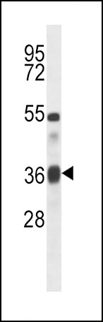
Western blot analysis in mouse heart tissue lysates (35ug/lane).Application
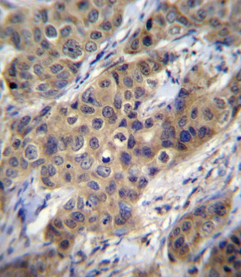
TGFB1 Antibody immunohistochemistry analysis in formalin fixed and paraffin embedded human breast carcinoma followed by peroxidase conjugation of the secondary antibody and DAB staining.Application
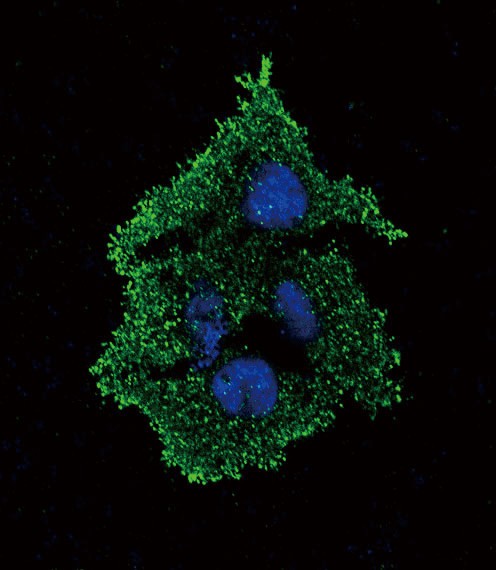
Confocal immunofluorescent analysis of TGFB1 Antibody with HepG2 cell followed by Alexa Fluor 488-conjugated goat anti-rabbit lgG (green). DAPI was used to stain the cell nuclear (blue).| Product Name | TGFB1 Antibody |
|---|---|
| Antibody Type | Primary Antibodies |
| Antigen Alias | Transforming growth factor beta-1, TGF-beta-1, Latency-associated peptide, LAP, TGFB1, TGFB |
| Product description | TGFB1 is a member of the transforming growth factor beta (TGFB) family of cytokines, which are multifunctional peptides that regulate proliferation, differentiation, adhesion, migration, and other functions in many cell types. Many cells have TGFB receptors, and the protein positively and negatively regulates many other growth factors. The secreted protein is cleaved into a latency-associated peptide (LAP) and a mature TGFB1 peptide, and is found in either a latent form composed of a TGFB1 homodimer, a LAP homodimer, and a latent TGFB1-binding protein, or in an active form composed of a TGFB1 homodimer. The mature peptide may also form heterodimers with other TGFB family members. This gene is frequently upregulated in tumor cells, and mutations in this gene result in Camurati-Engelmann disease.1) Perez, A.B., et al. Hum. Immunol. 71(11):1135-1140(2010) Xu, Z., et al. Biochem. Biophys. Res. Commun. 401(3):376-381(2010) Bran, G.M., et al. Anticancer Res. 30(9):3459-3463(2010) Zauli, G., et al. Blood 80(12):3036-3043(1992) Wrana, J.L., et al. Cell 71(6):1003-1014(1992) |
| Immunogen | This TGFB1 antibody is generated from rabbits immunized with a KLH conjugated synthetic peptide between 22-50 amino acids from the N-terminal region of human TGFB1. |
| Clonality | Polyclonal |
|---|---|
| Isotype | Ig |
| Host Species | Rabbit |
| Tested Applications | IFIHC-PWB |
| For WB starting dilution is: 1:1000 For IHC-P starting dilution is: 1:10~50 For IF starting dilution is: 1:10~50: |
|
| Species Reactivity | HumanMouse |
| Concentration | 1mg/ml |
| Purification | Affinity purified |
| Gene Symbol | TGFB1 |
|---|---|
| Alternative Names | Transforming growth factor beta-1 TGF-beta-1 Latency-associated peptide LAP TGFB1 TGFB |
| Molecular Weight(MW) | 44 kDa |
| Sequence Similarities | Predicted species reactivity based on immunogen sequence: Rat |
Application
Western Blot at 1:2000 dilution Lane 1: HepG2 whole cell lysate Lane 2: K562 whole cell lysate Lane 3: Raji whole cell lysate Lysates/proteins at 20 ug per lane.
Application
Western blot analysis in mouse heart tissue lysates (35ug/lane).
Application
TGFB1 Antibody immunohistochemistry analysis in formalin fixed and paraffin embedded human breast carcinoma followed by peroxidase conjugation of the secondary antibody and DAB staining.
Application
Confocal immunofluorescent analysis of TGFB1 Antibody with HepG2 cell followed by Alexa Fluor 488-conjugated goat anti-rabbit lgG (green). DAPI was used to stain the cell nuclear (blue).| Application Notes | For WB starting dilution is: 1:1000 For IHC-P starting dilution is: 1:10~50 For IF starting dilution is: 1:10~50: |
|---|
| Form | Liquid |
|---|---|
| Storage Instructions | Store at 4˚C for three months and -20˚C, stable for up to one year. As with all antibodies care should be taken to avoid repeated freeze thaw cycles. Antibodies should not be exposed to prolonged high temperatures. |
| Storage Buffer | Supplied in PBS with 0.09% (W/V) sodium azide. |


 Data sheet for OM293203