WB
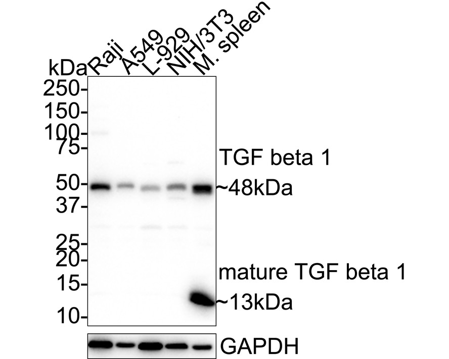
Western blot analysis of TGF beta 1 on different lysates with Rabbit anti-TGF beta 1 antibody at 1/1,000 dilution. Lane 1: Raji cell lysate, Lane 2: A549 cell lysate, Lane 3: L-929 cell lysate, Lane 4: NIH/3T3 cell lysate, Lane 5: Mouse spleen tissue lysate, Lysates/proteins at 20 µg/Lane. Exposure time: 2 minutes 50 seconds; 4-20% SDS-PAGE gel. Proteins were transferred to a PVDF membrane and blocked with 5% NFDM/TBST for 1 hour at room temperature. The primary antibody at 1/1,000 dilution was used in 5% NFDM/TBST at 4℃ overnight. Goat Anti-Rabbit IgG - HRP Secondary Antibody at 1/50,000 dilution was used for 1 hour at room temperature.IHC
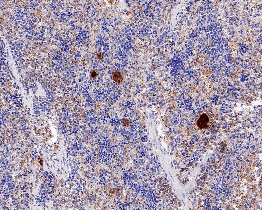
Immunohistochemical analysis of paraffin-embedded rat spleen tissue with Rabbit anti-TGF beta 1 antibody at 1/200 dilution. The section was pre-treated using heat mediated antigen retrieval with Tris-EDTA buffer (pH 9.0) for 20 minutes. The tissues were blocked in 1% BSA for 20 minutes at room temperature, washed with ddH2O and PBS, and then probed with the primary antibody at 1/200 dilution for 1 hour at room temperature. The detection was performed using an HRP conjugated compact polymer system. DAB was used as the chromogen. Tissues were counterstained with hematoxylin and mounted with DPX.| Product Name | TGF beta 1 Recombinant Rabbit Monoclonal Antibody |
|---|---|
| Antibody Type | Primary Antibodies |
| Immunogen | Synthetic peptide (KLH-coupled) corresponding to Human TGF-Beta 1 C-terminal. |
| Clonality | Monoclonal |
|---|---|
| Isotype | IgG |
| Host Species | Rabbit |
| Tested Applications | IHCWB |
| WB:1:1000-1:2000 IHC:1:200 |
|
| Species Reactivity | HumanMouseRat |
| Concentration | 1mg/ml |
| Purification | Protein A |
| Gene Symbol | TGFB1 |
|---|---|
| Gene Synonyms | CED LAP DPD1 TGFB IBDIMDE TGFbeta TGF-beta1 |
| Gene Full Name | transforming growth factor beta 1 |
| Gene Summary | This gene encodes a secreted ligand of the TGF-beta (transforming growth factor-beta) superfamily of proteins. Ligands of this family bind various TGF-beta receptors leading to recruitment and activation of SMAD family transcription factors that regulate gene expression. The encoded preproprotein is proteolytically processed to generate a latency-associated peptide (LAP) and a mature peptide, and is found in either a latent form composed of a mature peptide homodimer, a LAP homodimer, and a latent TGF-beta binding protein, or in an active form consisting solely of the mature peptide homodimer. The mature peptide may also form heterodimers with other TGFB family members. This encoded protein regulates cell proliferation, differentiation and growth, and can modulate expression and activation of other growth factors including interferon gamma and tumor necrosis factor alpha. This gene is frequently upregulated in tumor cells, and mutations in this gene result in Camurati-Engelmann disease. [provided by RefSeq, Aug 2016] |
| Molecular Weight(MW) | 44 kDa(Observed band size:48/13kDa) |
| Cellular Localization | Extracellular matrix, Secreted. |
WB
Western blot analysis of TGF beta 1 on different lysates with Rabbit anti-TGF beta 1 antibody at 1/1,000 dilution. Lane 1: Raji cell lysate, Lane 2: A549 cell lysate, Lane 3: L-929 cell lysate, Lane 4: NIH/3T3 cell lysate, Lane 5: Mouse spleen tissue lysate, Lysates/proteins at 20 µg/Lane. Exposure time: 2 minutes 50 seconds; 4-20% SDS-PAGE gel. Proteins were transferred to a PVDF membrane and blocked with 5% NFDM/TBST for 1 hour at room temperature. The primary antibody at 1/1,000 dilution was used in 5% NFDM/TBST at 4℃ overnight. Goat Anti-Rabbit IgG - HRP Secondary Antibody at 1/50,000 dilution was used for 1 hour at room temperature.
IHC
Immunohistochemical analysis of paraffin-embedded rat spleen tissue with Rabbit anti-TGF beta 1 antibody at 1/200 dilution. The section was pre-treated using heat mediated antigen retrieval with Tris-EDTA buffer (pH 9.0) for 20 minutes. The tissues were blocked in 1% BSA for 20 minutes at room temperature, washed with ddH2O and PBS, and then probed with the primary antibody at 1/200 dilution for 1 hour at room temperature. The detection was performed using an HRP conjugated compact polymer system. DAB was used as the chromogen. Tissues were counterstained with hematoxylin and mounted with DPX.| Application Notes | WB:1:1000-1:2000 IHC:1:200 |
|---|
| Form | Liquid |
|---|---|
| Storage Instructions | Store at +4℃ after thawing. Aliquot store at -20℃. Avoid repeated freeze / thaw cycles. |
| Storage Buffer | PBS (pH7.4), 0.1% BSA, 40% Glycerol. Preservative: 0.05% Sodium Azide. |


 Data sheet for OM643272