WB
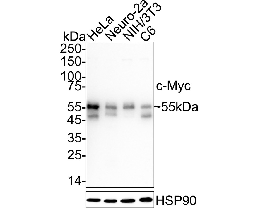
Western blot analysis of c-Myc on different lysates with Rabbit anti-c-Myc antibody at 1/1,000 dilution. Lane 1: HeLa cell lysate, Lane 2: Neuro-2a cell lysate, Lane 3: NIH/3T3 cell lysate, Lane 4: C6 cell lysate, Lysates/proteins at 20 µg/Lane. Exposure time: 1 minute; 4-20% SDS-PAGE gel. Proteins were transferred to a PVDF membrane and blocked with 5% NFDM/TBST for 1 hour at room temperature. The primary antibody at 1/1,000 dilution was used in 5% NFDM/TBST at 4℃ overnight. Goat Anti-Rabbit IgG - HRP Secondary Antibody at 1/50,000 dilution was used for 1 hour at room temperature.IHC
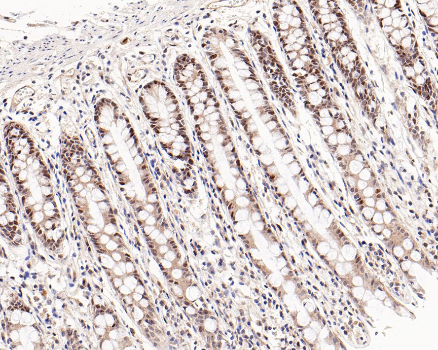
Immunohistochemical analysis of paraffin-embedded human colon tissue with Rabbit anti-c-Myc antibody at 1/200 dilution. The section was pre-treated using heat mediated antigen retrieval with sodium citrate buffer (pH 6.0) for 2 minutes. The tissues were blocked in 1% BSA for 20 minutes at room temperature, washed with ddH2O and PBS, and then probed with the primary antibody at 1/200 dilution for 1 hour at room temperature. The detection was performed using an HRP conjugated compact polymer system. DAB was used as the chromogen. Tissues were counterstained with hematoxylin and mounted with DPX.IHC
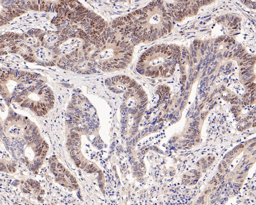
Immunohistochemical analysis of paraffin-embedded human colon carcinoma tissue with Rabbit anti-c-Myc antibody at 1/200 dilution. The section was pre-treated using heat mediated antigen retrieval with sodium citrate buffer (pH 6.0) for 2 minutes. The tissues were blocked in 1% BSA for 20 minutes at room temperature, washed with ddH2O and PBS, and then probed with the primary antibody at 1/200 dilution for 1 hour at room temperature. The detection was performed using an HRP conjugated compact polymer system. DAB was used as the chromogen. Tissues were counterstained with hematoxylin and mounted with DPX.IP
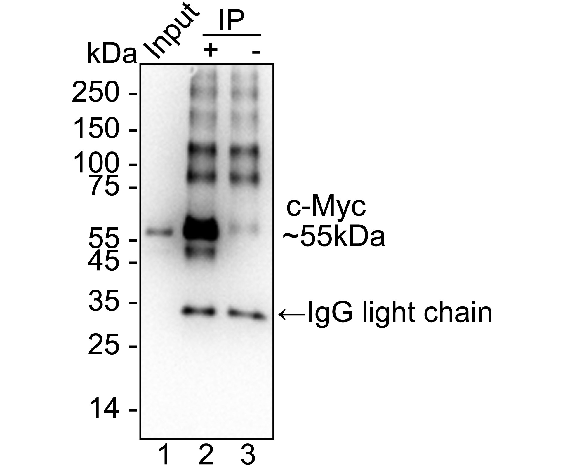
c-Myc was immunoprecipitated from 0.2 mg HeLa cell lysate with Rabbit anti-c-Myc antibody at 2 µg/25 µl agarose. Western blot was performed from the immunoprecipitate using Rabbit anti-c-Myc antibody at 1/1,000 dilution. Anti-Rabbit IgG for IP Nano-secondary antibody at 1/5,000 dilution was used for 1 hour at room temperature. Lane 1: HeLa cell lysate (input), Lane 2: Rabbit anti-c-Myc antibody IP in HeLa cell lysate, Lane 3: Rabbit IgG instead of Rabbit anti-c-Myc antibody in HeLa cell lysate, Blocking/Dilution buffer: 5% NFDM/TBST, Exposure time: 30 seconds.| Product Name | c-Myc Recombinant Rabbit Monoclonal Antibody |
|---|---|
| Antibody Type | Primary Antibodies |
| Immunogen | Synthetic peptide. |
| Clonality | monoclonal |
|---|---|
| Isotype | IgG |
| Host Species | Rabbit |
| Tested Applications | IHCIPWB |
| WB:1:1000 IHC:1:200 IP:1-2μg/sample |
|
| Species Reactivity | HumanMouseRat |
| Concentration | 1mg/ml |
| Purification | Protein A |
| Gene Symbol | MYC |
|---|---|
| Gene Synonyms | MRTL MYCC c-Myc bHLHe39 |
| Gene Full Name | MYC proto-oncogene, bHLH transcription factor |
| Gene Summary | This gene is a proto-oncogene and encodes a nuclear phosphoprotein that plays a role in cell cycle progression, apoptosis and cellular transformation. The encoded protein forms a heterodimer with the related transcription factor MAX. This complex binds to the E box DNA consensus sequence and regulates the transcription of specific target genes. Amplification of this gene is frequently observed in numerous human cancers. Translocations involving this gene are associated with Burkitt lymphoma and multiple myeloma in human patients. There is evidence to show that translation initiates both from an upstream, in-frame non-AUG (CUG) and a downstream AUG start site, resulting in the production of two isoforms with distinct N-termini. [provided by RefSeq, Aug 2017] |
| Molecular Weight(MW) | 49kDa(Observed band size: 55kDa) |
| Cellular Localization | Nucleoplasm, nucleolus. |
WB
Western blot analysis of c-Myc on different lysates with Rabbit anti-c-Myc antibody at 1/1,000 dilution. Lane 1: HeLa cell lysate, Lane 2: Neuro-2a cell lysate, Lane 3: NIH/3T3 cell lysate, Lane 4: C6 cell lysate, Lysates/proteins at 20 µg/Lane. Exposure time: 1 minute; 4-20% SDS-PAGE gel. Proteins were transferred to a PVDF membrane and blocked with 5% NFDM/TBST for 1 hour at room temperature. The primary antibody at 1/1,000 dilution was used in 5% NFDM/TBST at 4℃ overnight. Goat Anti-Rabbit IgG - HRP Secondary Antibody at 1/50,000 dilution was used for 1 hour at room temperature.
IHC
Immunohistochemical analysis of paraffin-embedded human colon tissue with Rabbit anti-c-Myc antibody at 1/200 dilution. The section was pre-treated using heat mediated antigen retrieval with sodium citrate buffer (pH 6.0) for 2 minutes. The tissues were blocked in 1% BSA for 20 minutes at room temperature, washed with ddH2O and PBS, and then probed with the primary antibody at 1/200 dilution for 1 hour at room temperature. The detection was performed using an HRP conjugated compact polymer system. DAB was used as the chromogen. Tissues were counterstained with hematoxylin and mounted with DPX.
IHC
Immunohistochemical analysis of paraffin-embedded human colon carcinoma tissue with Rabbit anti-c-Myc antibody at 1/200 dilution. The section was pre-treated using heat mediated antigen retrieval with sodium citrate buffer (pH 6.0) for 2 minutes. The tissues were blocked in 1% BSA for 20 minutes at room temperature, washed with ddH2O and PBS, and then probed with the primary antibody at 1/200 dilution for 1 hour at room temperature. The detection was performed using an HRP conjugated compact polymer system. DAB was used as the chromogen. Tissues were counterstained with hematoxylin and mounted with DPX.
IP
c-Myc was immunoprecipitated from 0.2 mg HeLa cell lysate with Rabbit anti-c-Myc antibody at 2 µg/25 µl agarose. Western blot was performed from the immunoprecipitate using Rabbit anti-c-Myc antibody at 1/1,000 dilution. Anti-Rabbit IgG for IP Nano-secondary antibody at 1/5,000 dilution was used for 1 hour at room temperature. Lane 1: HeLa cell lysate (input), Lane 2: Rabbit anti-c-Myc antibody IP in HeLa cell lysate, Lane 3: Rabbit IgG instead of Rabbit anti-c-Myc antibody in HeLa cell lysate, Blocking/Dilution buffer: 5% NFDM/TBST, Exposure time: 30 seconds.| Application Notes | WB:1:1000 IHC:1:200 IP:1-2μg/sample |
|---|
| Form | Liquid |
|---|---|
| Storage Instructions | Store at +4℃ after thawing. Aliquot store at -20℃. Avoid repeated freeze / thaw cycles. |
| Storage Buffer | PBS (pH7.4), 0.1% BSA, 40% Glycerol. Preservative: 0.05% Sodium Azide. |


 Data sheet for OM644179