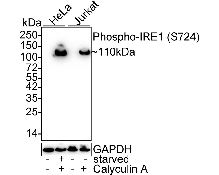

WB
Western blot analysis of Phospho-IRE1 (S724) on different lysates with Rabbit anti-Phospho-IRE1 (S724) antibody at 1/1,000 dilution. Lane 1: HeLa cell lysate (20 µg/Lane), Lane 2: HeLa starved for 3 hours then treated with 100nM Calyculin A for 30 minutes cell lysate (20 µg/Lane), Lane 3: Jurkat cell lysate (20 µg/Lane), Lane 4: Jurkat treated with 100nM Calyculin A for 30 minutes cell lysate (20 µg/Lane), Exposure time: 5 minutes 10 seconds; 4-20% SDS-PAGE gel. Proteins were transferred to a PVDF membrane and blocked with 5% NFDM/TBST for 1 hour at room temperature. The primary antibody at 1/1,000 dilution was used in 5% NFDM/TBST at 4℃ overnight. Goat Anti-Rabbit IgG - HRP Secondary Antibody at 1/50,000 dilution was used for 1 hour at room temperature.IHC
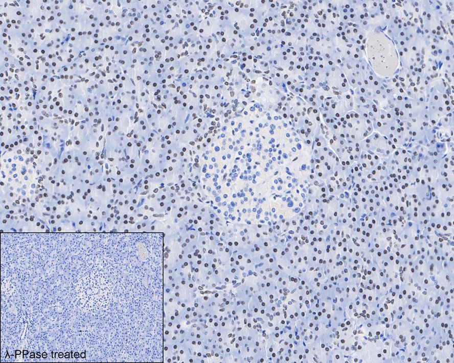
Immunohistochemical analysis of paraffin-embedded human pancreas tissue untreated / treated with λpp with Rabbit anti-Phospho-IRE1 (S724) antibody at 1/500 dilution. The section was pre-treated using heat mediated antigen retrieval with Tris-EDTA buffer (pH 9.0) for 20 minutes. The tissues were blocked in 1% BSA for 20 minutes at room temperature, washed with ddH2O and PBS, and then probed with the primary antibody at 1/500 dilution for 1 hour at room temperature. The detection was performed using an HRP conjugated compact polymer system. DAB was used as the chromogen. Tissues were counterstained with hematoxylin and mounted with DPX.ICC/IF
Immunocytochemistry analysis of Jurkat cells treated with 100nM Calyculin A for 30 minutes labeling Phospho-IRE1 (S724) with Rabbit anti-Phospho-IRE1 (S724) antibody at 1/500 dilution. Cells were fixed in 4% paraformaldehyde for 20 minutes at room temperature, permeabilized with 0.1% Triton X-100 in PBS for 5 minutes at room temperature, then blocked with 1% BSA in 10% negative goat serum for 1 hour at room temperature. Cells were then incubated with Rabbit anti-Phospho-IRE1 (S724) antibody at 1/500 dilution in 1% BSA in PBST overnight at 4 ℃. Goat Anti-Rabbit IgG H&L (488) was used as the secondary antibody at 1/1,000 dilution. PBS instead of the primary antibody was used as the secondary antibody only control. Nuclear DNA was labelled in blue with DAPI. Beta tubulin (red) was stained at 1/100 dilution overnight at +4℃. Goat Anti-Mouse IgG H&L (594) was used as the secondary antibody at 1/1,000 dilution.FC
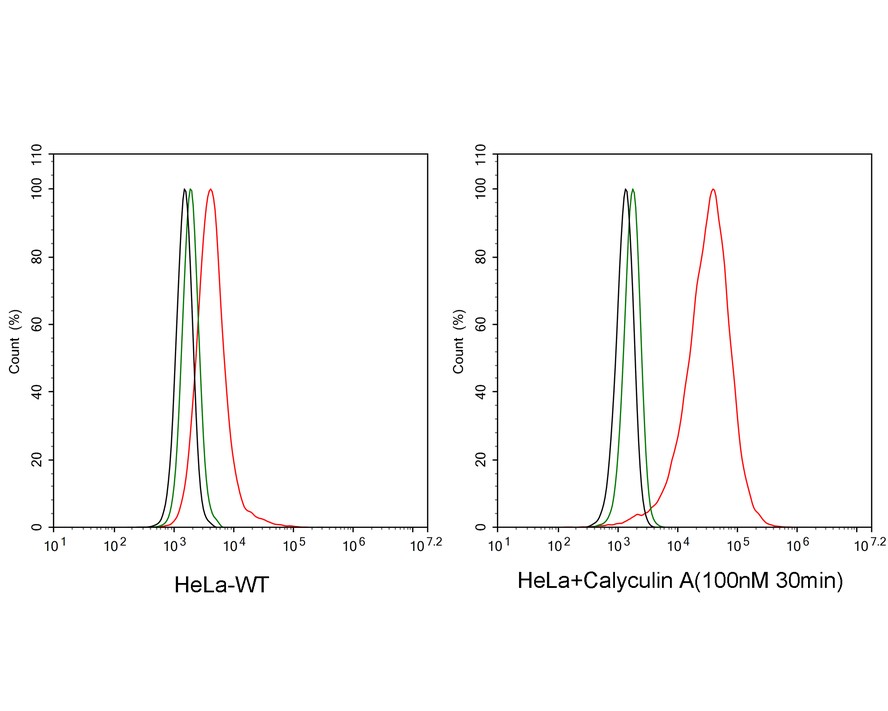
Flow cytometric analysis of HeLa cells treated with or without 100nM Calyculin A for 30 minutes labeling Phospho-IRE1 (S724). Cells were fixed and permeabilized. Then stained with the primary antibody (1μg/mL) (red) compared with Rabbit IgG Isotype Control (green). After incubation of the primary antibody at +4℃ for an hour, the cells were stained with a iFluor™ 488 conjugate-Goat anti-Rabbit IgG Secondary antibody at 1/1,000 dilution for 30 minutes at +4℃. Unlabelled sample was used as a control (cells without incubation with primary antibody; black).| Product Name | Phospho-IRE1 (S724) Recombinant Rabbit Monoclonal Antibody |
|---|---|
| Antibody Type | Primary Antibodies |
| Immunogen | Synthetic phospho-peptide corresponding to residues surrounding Ser724 of human IRE1. |
| Clonality | monoclonal |
|---|---|
| Isotype | IgG |
| Host Species | Rabbit |
| Tested Applications | FCICC/IFIHCWB |
| WB:1:1000 IHC:1:500 ICC/IF:1:500 FC:1:1000 |
|
| Species Reactivity | HumanMouse |
| Concentration | 1mg/ml |
| Purification | Protein A |
| Gene Symbol | ERN1 |
|---|---|
| Gene Synonyms | IRE1 IRE1P IRE1a hIRE1p |
| Gene Full Name | endoplasmic reticulum to nucleus signaling 1 |
| Gene Summary | This gene encodes the transmembrane protein kinase inositol-requiring enzyme 1. The encoded protein contains two functional catalytic domains, a serine/threonine-protein kinase domain and an endoribonuclease domain. This protein functions as a sensor of unfolded proteins in the endoplasmic reticulum (ER) and triggers an intracellular signaling pathway termed the unfolded protein response (UPR). The UPR is an ER stress response that is conserved from yeast to mammals and activates genes involved in degrading misfolded proteins, regulating protein synthesis and activating molecular chaperones. This protein specifically mediates the splicing and activation of the stress response transcription factor X-box binding protein 1. [provided by RefSeq, Aug 2017] |
| Molecular Weight(MW) | 110kDa |
| Cellular Localization | Endoplasmic reticulum membrane; nucleus. |
WB
Western blot analysis of Phospho-IRE1 (S724) on different lysates with Rabbit anti-Phospho-IRE1 (S724) antibody at 1/1,000 dilution. Lane 1: HeLa cell lysate (20 µg/Lane), Lane 2: HeLa starved for 3 hours then treated with 100nM Calyculin A for 30 minutes cell lysate (20 µg/Lane), Lane 3: Jurkat cell lysate (20 µg/Lane), Lane 4: Jurkat treated with 100nM Calyculin A for 30 minutes cell lysate (20 µg/Lane), Exposure time: 5 minutes 10 seconds; 4-20% SDS-PAGE gel. Proteins were transferred to a PVDF membrane and blocked with 5% NFDM/TBST for 1 hour at room temperature. The primary antibody at 1/1,000 dilution was used in 5% NFDM/TBST at 4℃ overnight. Goat Anti-Rabbit IgG - HRP Secondary Antibody at 1/50,000 dilution was used for 1 hour at room temperature.
IHC
Immunohistochemical analysis of paraffin-embedded human pancreas tissue untreated / treated with λpp with Rabbit anti-Phospho-IRE1 (S724) antibody at 1/500 dilution. The section was pre-treated using heat mediated antigen retrieval with Tris-EDTA buffer (pH 9.0) for 20 minutes. The tissues were blocked in 1% BSA for 20 minutes at room temperature, washed with ddH2O and PBS, and then probed with the primary antibody at 1/500 dilution for 1 hour at room temperature. The detection was performed using an HRP conjugated compact polymer system. DAB was used as the chromogen. Tissues were counterstained with hematoxylin and mounted with DPX.
ICC/IF
Immunocytochemistry analysis of Jurkat cells treated with 100nM Calyculin A for 30 minutes labeling Phospho-IRE1 (S724) with Rabbit anti-Phospho-IRE1 (S724) antibody at 1/500 dilution. Cells were fixed in 4% paraformaldehyde for 20 minutes at room temperature, permeabilized with 0.1% Triton X-100 in PBS for 5 minutes at room temperature, then blocked with 1% BSA in 10% negative goat serum for 1 hour at room temperature. Cells were then incubated with Rabbit anti-Phospho-IRE1 (S724) antibody at 1/500 dilution in 1% BSA in PBST overnight at 4 ℃. Goat Anti-Rabbit IgG H&L (488) was used as the secondary antibody at 1/1,000 dilution. PBS instead of the primary antibody was used as the secondary antibody only control. Nuclear DNA was labelled in blue with DAPI. Beta tubulin (red) was stained at 1/100 dilution overnight at +4℃. Goat Anti-Mouse IgG H&L (594) was used as the secondary antibody at 1/1,000 dilution.
FC
Flow cytometric analysis of HeLa cells treated with or without 100nM Calyculin A for 30 minutes labeling Phospho-IRE1 (S724). Cells were fixed and permeabilized. Then stained with the primary antibody (1μg/mL) (red) compared with Rabbit IgG Isotype Control (green). After incubation of the primary antibody at +4℃ for an hour, the cells were stained with a iFluor™ 488 conjugate-Goat anti-Rabbit IgG Secondary antibody at 1/1,000 dilution for 30 minutes at +4℃. Unlabelled sample was used as a control (cells without incubation with primary antibody; black).| Application Notes | WB:1:1000 IHC:1:500 ICC/IF:1:500 FC:1:1000 |
|---|
| Form | Liquid |
|---|---|
| Storage Instructions | Store at +4℃ after thawing. Aliquot store at -20℃. Avoid repeated freeze / thaw cycles. |
| Storage Buffer | 1*TBS (pH7.4), 0.05% BSA, 40% Glycerol. Preservative: 0.05% Sodium Azide. |


 Data sheet for OM644188