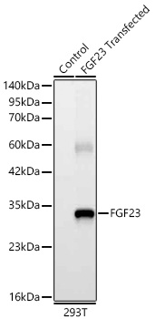
WB
Western blot analysis of lysates from wild type (WT) and 293T cells transfected with FGF23 using FGF23 Rabbit mAb at 1:6000 dilution. Secondary antibody: HRP Goat Anti-Rabbit IgG (H+L)at 1:10000 dilution. Lysates/proteins: 25ug per lane. Blocking buffer: 3% nonfat dry milk in TBST. Detection: ECL Basic Kit. Exposure time: 5s.ICC/IF
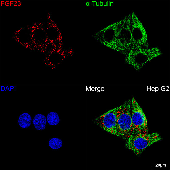
Confocal imaging of Hep G2 cells using FGF23 Rabbit mAb (dilution 1:200) followed by a further incubation with Cy3 Goat Anti-Rabbit IgG (H+L) (dilution 1:500) (Red). The cells were counterstained with α-Tubulin Mouse mAb (dilution 1:400) followed by incubation with Omnimabs® 488 Goat Anti-Mouse IgG(H&L)(dilution 1:500)(Green). DAPI was used for nuclear staining (Blue). Objective: 100x.| Product Name | FGF23 Rabbit mAb |
|---|---|
| Antibody Type | Primary Antibodies |
| Immunogen | Recombinant fusion protein containing a sequence corresponding to amino acids 25-251 of human FGF23 (NP_065689.1) |
| Clonality | monoclonal |
|---|---|
| Isotype | IgG |
| Host Species | Rabbit |
| Tested Applications | ICC/IFWB |
| WB:1:2000-1:10000 ICC/IF:1:50-1:200 |
|
| Species Reactivity | Human |
| Concentration | 1mg/ml |
| Purification | Affinity purified |
| Gene Symbol | FGF23 |
|---|---|
| Gene Synonyms | ADHR FGFN HYPF HFTC2 HPDR2 PHPTC |
| Gene Full Name | fibroblast growth factor 23 |
| Gene Summary | This gene encodes a member of the fibroblast growth factor family of proteins, which possess broad mitogenic and cell survival activities and are involved in a variety of biological processes. The product of this gene regulates phosphate homeostasis and transport in the kidney. The full-length, functional protein may be deactivated via cleavage into N-terminal and C-terminal chains. Mutation of this cleavage site causes autosomal dominant hypophosphatemic rickets (ADHR). Mutations in this gene are also associated with hyperphosphatemic familial tumoral calcinosis (HFTC). [provided by RefSeq, Feb 2013] |
| Molecular Weight(MW) | 28kDa |
| Cellular Localization | Secreted. |
WB
Western blot analysis of lysates from wild type (WT) and 293T cells transfected with FGF23 using FGF23 Rabbit mAb at 1:6000 dilution. Secondary antibody: HRP Goat Anti-Rabbit IgG (H+L)at 1:10000 dilution. Lysates/proteins: 25ug per lane. Blocking buffer: 3% nonfat dry milk in TBST. Detection: ECL Basic Kit. Exposure time: 5s.
ICC/IF
Confocal imaging of Hep G2 cells using FGF23 Rabbit mAb (dilution 1:200) followed by a further incubation with Cy3 Goat Anti-Rabbit IgG (H+L) (dilution 1:500) (Red). The cells were counterstained with α-Tubulin Mouse mAb (dilution 1:400) followed by incubation with Omnimabs® 488 Goat Anti-Mouse IgG(H&L)(dilution 1:500)(Green). DAPI was used for nuclear staining (Blue). Objective: 100x.| Application Notes | WB:1:2000-1:10000 ICC/IF:1:50-1:200 |
|---|
| Form | Liquid |
|---|---|
| Storage Instructions | Store at -20℃. Avoid freeze / thaw cycles. |
| Storage Buffer | Buffer: PBS with 0.05% proclin300, 0.05% BSA, 50% glycerol, pH7.3. |


 Data sheet for OM644231