Application
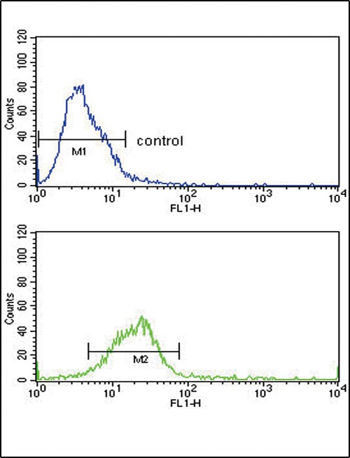
Flow cytometric analysis of Hela cells (bottom histogram) compared to a negative control cell (top histogram).FITC-conjugated goat-anti-rabbit secondary antibodies were used for the analysis.Application
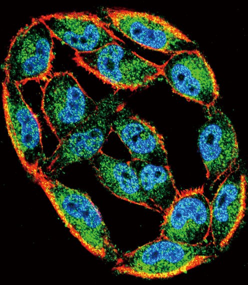
Confocal immunofluorescent analysis of HSPA8 Antibody with A2058 cell followed by Alexa Fluor 488-conjugated goat anti-rabbit lgG (green). Actin filaments have been labeled with Alexa Fluor 555 phalloidin (red).DAPI was used to stain the cell nuclear (blue).Application
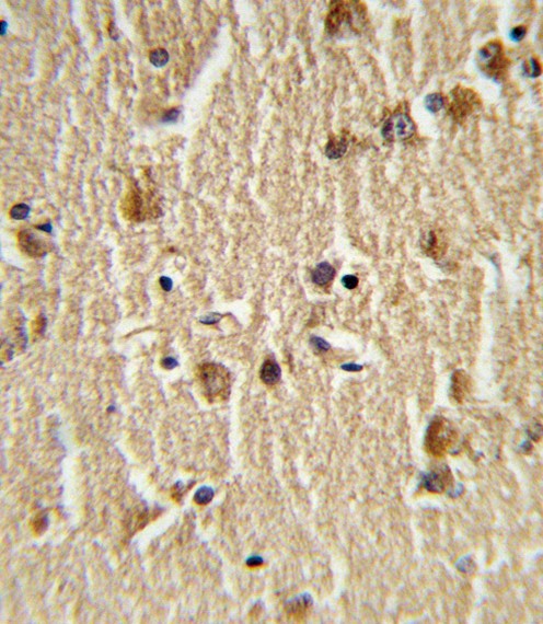
Formalin-fixed and paraffin-embedded human brain tissue reacted with HSPA8 Antibody (N-term), which was peroxidase-conjugated to the secondary antibody, followed by DAB staining.Application
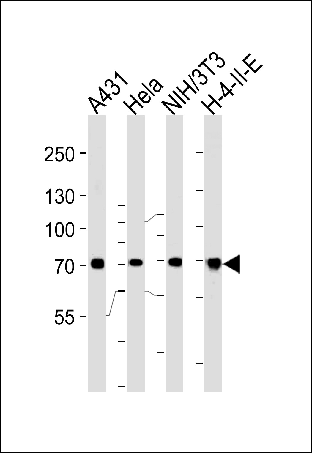
Western blot analysis of lysates from A431,Hela,mouse NIH/3T3,H-4-II-E cell line (from left to right),using HSPA8 Antibody .AP2872a was diluted at 1:1000 at each lane.| Product Name | HSPA8 Antibody |
|---|---|
| Antibody Type | Primary Antibodies |
| Antigen Alias | Heat shock cognate 71 kDa protein, Heat shock 70 kDa protein 8, Lipopolysaccharide-associated protein 1, LAP-1, LPS-associated protein 1, HSPA8, HSC70, HSP73, HSPA10 |
| Product description | HSPA8 belongs to the heat shock protein 70 family which contains both heat-inducible and constitutively expressed members. The latter are called heat-shock cognate proteins. HSPA8 is a heat-shock cognate protein. This protein binds to nascent polypeptides to facilitate correct folding. The protein also functions as an ATPase in the disassembly of clathrin-coated vesicles during transport of membrane components through the cell.1) Tsukahara F., Yoshioka T.Mol. Pharmacol. 58:1257-1263(2000) |
| Immunogen | This HSPA8 antibody is generated from rabbits immunized with a KLH conjugated synthetic peptide between 82-110 amino acids from the N-terminal region of human HSPA8. |
| Clonality | Polyclonal |
|---|---|
| Isotype | Ig |
| Host Species | Rabbit |
| Tested Applications | FACSIFIHC-PWB |
| For WB starting dilution is: 1:1000 For IHC-P starting dilution is: 1:10~50 For IF starting dilution is: 1:10~50 For FACS starting dilution is: 1:10~50: |
|
| Species Reactivity | Human |
| Concentration | 1mg/ml |
| Gene Symbol | HSPA8 |
|---|---|
| Alternative Names | Heat shock cognate 71 kDa protein Heat shock 70 kDa protein 8 Lipopolysaccharide-associated protein 1 LAP-1 LPS-associated protein 1 HSPA8 HSC70 HSP73 HSPA10 |
| Molecular Weight(MW) | 71 kDa |
| Sequence Similarities | Predicted species reactivity based on immunogen sequence: Bovine |
Application
Flow cytometric analysis of Hela cells (bottom histogram) compared to a negative control cell (top histogram).FITC-conjugated goat-anti-rabbit secondary antibodies were used for the analysis.
Application
Confocal immunofluorescent analysis of HSPA8 Antibody with A2058 cell followed by Alexa Fluor 488-conjugated goat anti-rabbit lgG (green). Actin filaments have been labeled with Alexa Fluor 555 phalloidin (red).DAPI was used to stain the cell nuclear (blue).
Application
Formalin-fixed and paraffin-embedded human brain tissue reacted with HSPA8 Antibody (N-term), which was peroxidase-conjugated to the secondary antibody, followed by DAB staining.
Application
Western blot analysis of lysates from A431,Hela,mouse NIH/3T3,H-4-II-E cell line (from left to right),using HSPA8 Antibody .AP2872a was diluted at 1:1000 at each lane.| Application Notes | For WB starting dilution is: 1:1000 For IHC-P starting dilution is: 1:10~50 For IF starting dilution is: 1:10~50 For FACS starting dilution is: 1:10~50: |
|---|
| Form | Liquid |
|---|---|
| Storage Instructions | Store at 4˚C for three months and -20˚C, stable for up to one year. As with all antibodies care should be taken to avoid repeated freeze thaw cycles. Antibodies should not be exposed to prolonged high temperatures. |
| Storage Buffer | Supplied in PBS with 0.09% (W/V) sodium azide. |


 Data sheet for OM281434