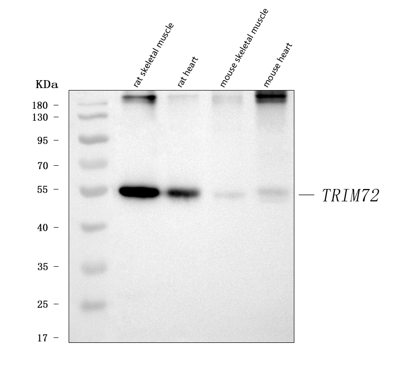


WB
Western blot analysis of TRIM72 using anti-TRIM72 antibody. The sample well of each lane was loaded with 30 ug of sample under reducing conditions. Lane 1: rat skeletal muscle tissue lysates, Lane 2: rat heart tissue lysates, Lane 3: mouse skeletal muscle tissue lysates, Lane 4: mouse heart tissue lysates. After electrophoresis, proteins were transferred to a membrane. Then the membrane was incubated with rabbit anti-TRIM72 antigen affinity purified polyclonal antibody at a dilution of 1:1000 and probed with a goat anti-rabbit IgG-HRP secondary antibody. The signal is developed using ECL Plus Western Blotting Substrate.IHC
IHC analysis of TRIM72 using anti-TRIM72 antibody. TRIM72 was detected in a paraffin-embedded section of human skeletal muscle tissue. The tissue section was incubated with rabbit anti-TRIM72 Antibody at a dilution of 1:200 and developed using HRP Conjugated Rabbit IgG Super Vision Assay Kit with DAB as the chromogen.IF-P
IF analysis using anti-TRIM72 antibody. detected in paraffin-embedded section of human skeletal tissue. The tissue section were stained using the Dylight550-conjugated Anti-rabbit IgG Secondary Antibody (red) and counterstained with DAPI (blue).FC
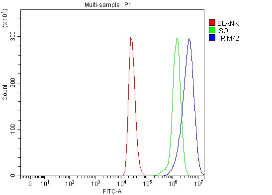
Flow Cytometry analysis of THP-1 cells using anti-TRIM72 antibody. Overlay histogram showing THP-1 cells stained with anti-TRIM72 antibody (Blue line). To facilitate intracellular staining, cells were fixed with 4% paraformaldehyde and permeabilized with permeabilization buffer. The cells were blocked with 10% normal goat serum. And then incubated with rabbit anti-TRIM72 Antibody at 1:100 dilution for 30 min at 20°C. DyLight®488 conjugated goat anti-rabbit IgG was used as secondary antibody at 1:100 dilution for 30 minutes at 20°C. Isotype control antibody (Green line) was rabbit IgG at 1:100 dilution used under the same conditions. Unlabelled sample without incubation with primary antibody and secondary antibody (Red line) was used as a blank control.| Product Name | Rabbit polyclonal antibody to TRIM72 |
|---|---|
| Antibody Type | Primary Antibodies |
| Immunogen | E. coli-derived human MG53/TRIM72 recombinant protein (Position: E28-A477). Human TRIM72 shares 90.9% and 90.7% amino acid (aa) sequence identity with mouse and rat TRIM72, respectively. |
| Clonality | polyclonal |
|---|---|
| Isotype | IgG |
| Host Species | Rabbit |
| Tested Applications | FCIF-PIHCWB |
| WB:1:500-1:2000 IHC:1:50-1:400 IF-P:1:50-1:400 FC:1:50-1:200 |
|
| Species Reactivity | HumanMouseRat |
| Concentration | 0.5mg/ml |
| Purification | Affinity purified |
| Gene Symbol | TRIM72 |
|---|---|
| Gene Synonyms | MG53 |
| Gene Full Name | tripartite motif containing 72 |
| Gene Summary | Enables identical protein binding activity. Predicted to be involved in several processes, including plasma membrane repair; proteasome-mediated ubiquitin-dependent protein catabolic process; and protein homooligomerization. Predicted to act upstream of or within negative regulation of insulin receptor signaling pathway; negative regulation of insulin-like growth factor receptor signaling pathway; and negative regulation of myotube differentiation. Predicted to be located in cytoplasmic vesicle membrane. Predicted to be active in cytoplasm and sarcolemma. [provided by Alliance of Genome Resources, Feb 2025] |
| Molecular Weight(MW) | 53kDa |
| Cellular Localization | Cell membrane,Cytoplasmic vesicle,Membrane. |
WB
Western blot analysis of TRIM72 using anti-TRIM72 antibody. The sample well of each lane was loaded with 30 ug of sample under reducing conditions. Lane 1: rat skeletal muscle tissue lysates, Lane 2: rat heart tissue lysates, Lane 3: mouse skeletal muscle tissue lysates, Lane 4: mouse heart tissue lysates. After electrophoresis, proteins were transferred to a membrane. Then the membrane was incubated with rabbit anti-TRIM72 antigen affinity purified polyclonal antibody at a dilution of 1:1000 and probed with a goat anti-rabbit IgG-HRP secondary antibody. The signal is developed using ECL Plus Western Blotting Substrate.
IHC
IHC analysis of TRIM72 using anti-TRIM72 antibody. TRIM72 was detected in a paraffin-embedded section of human skeletal muscle tissue. The tissue section was incubated with rabbit anti-TRIM72 Antibody at a dilution of 1:200 and developed using HRP Conjugated Rabbit IgG Super Vision Assay Kit with DAB as the chromogen.
IF-P
IF analysis using anti-TRIM72 antibody. detected in paraffin-embedded section of human skeletal tissue. The tissue section were stained using the Dylight550-conjugated Anti-rabbit IgG Secondary Antibody (red) and counterstained with DAPI (blue).
FC
Flow Cytometry analysis of THP-1 cells using anti-TRIM72 antibody. Overlay histogram showing THP-1 cells stained with anti-TRIM72 antibody (Blue line). To facilitate intracellular staining, cells were fixed with 4% paraformaldehyde and permeabilized with permeabilization buffer. The cells were blocked with 10% normal goat serum. And then incubated with rabbit anti-TRIM72 Antibody at 1:100 dilution for 30 min at 20°C. DyLight®488 conjugated goat anti-rabbit IgG was used as secondary antibody at 1:100 dilution for 30 minutes at 20°C. Isotype control antibody (Green line) was rabbit IgG at 1:100 dilution used under the same conditions. Unlabelled sample without incubation with primary antibody and secondary antibody (Red line) was used as a blank control.| Application Notes | WB:1:500-1:2000 IHC:1:50-1:400 IF-P:1:50-1:400 FC:1:50-1:200 |
|---|
| Form | Liquid |
|---|---|
| Storage Instructions | 12 months from date of receipt, -20℃ as supplied. 6 months 2 to 8℃ after reconstitution. Avoid repeated freezing and thawing. |
| Storage Buffer | 500ug/ml antibody with PBS, 0.02% NaN3, 1 mg/ml BSA and 50% glycerol. |


 Data sheet for OM643980