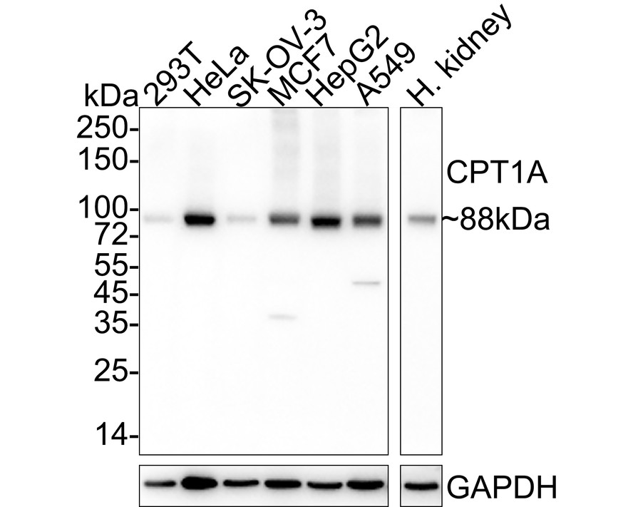
WB
Western blot analysis of CPT1A on different lysates with Rabbit anti-CPT1A antibody at 1/2,000 dilution. Lane 1: 293T cell lysate, Lane 2: HeLa cell lysate, Lane 3: SK-OV-3 cell lysate, Lane 4: MCF7 cell lysate, Lane 5: HepG2 cell lysate, Lane 6: A549 cell lysate, Lane 7: Human kidney tissue lysate, Lysates/proteins at 20 µg/Lane. Exposure time: 30 seconds; 4-20% SDS-PAGE gel. Proteins were transferred to a PVDF membrane and blocked with 5% NFDM/TBST for 1 hour at room temperature. The primary antibody at 1/2,000 dilution was used in 5% NFDM/TBST at 4℃ overnight. Goat Anti-Rabbit IgG - HRP Secondary Antibody at 1/50,000 dilution was used for 1 hour at room temperature.IHC
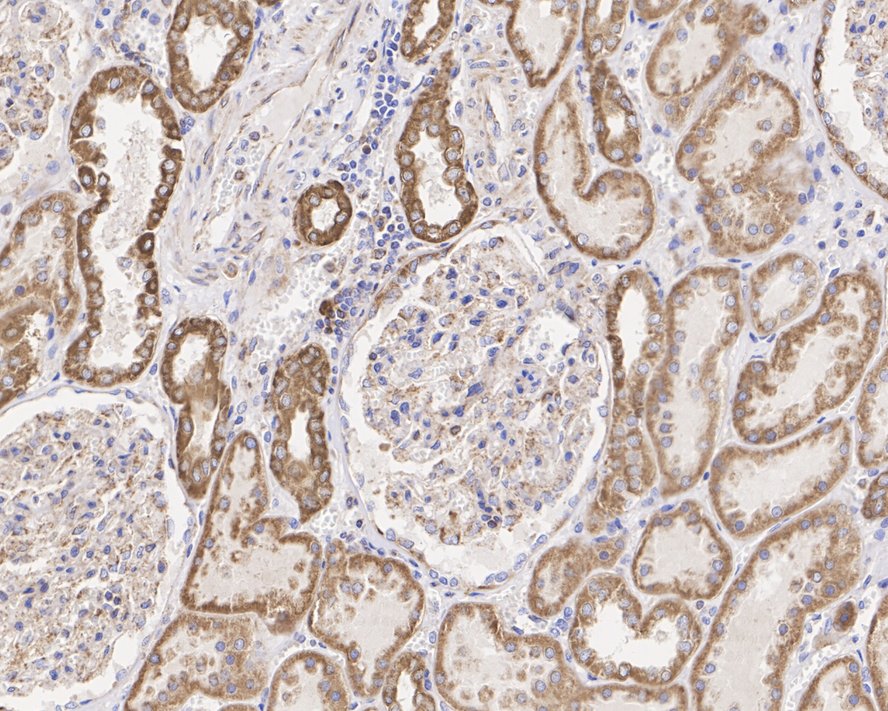
Immunohistochemical analysis of paraffin-embedded human kidney tissue with Rabbit anti-CPT1A antibody at 1/2,000 dilution. The section was pre-treated using heat mediated antigen retrieval with Tris-EDTA buffer (pH 9.0) for 20 minutes. The tissues were blocked in 1% BSA for 20 minutes at room temperature, washed with ddH2O and PBS, and then probed with the primary antibody at 1/2,000 dilution for 1 hour at room temperature. The detection was performed using an HRP conjugated compact polymer system. DAB was used as the chromogen. Tissues were counterstained with hematoxylin and mounted with DPX.IP
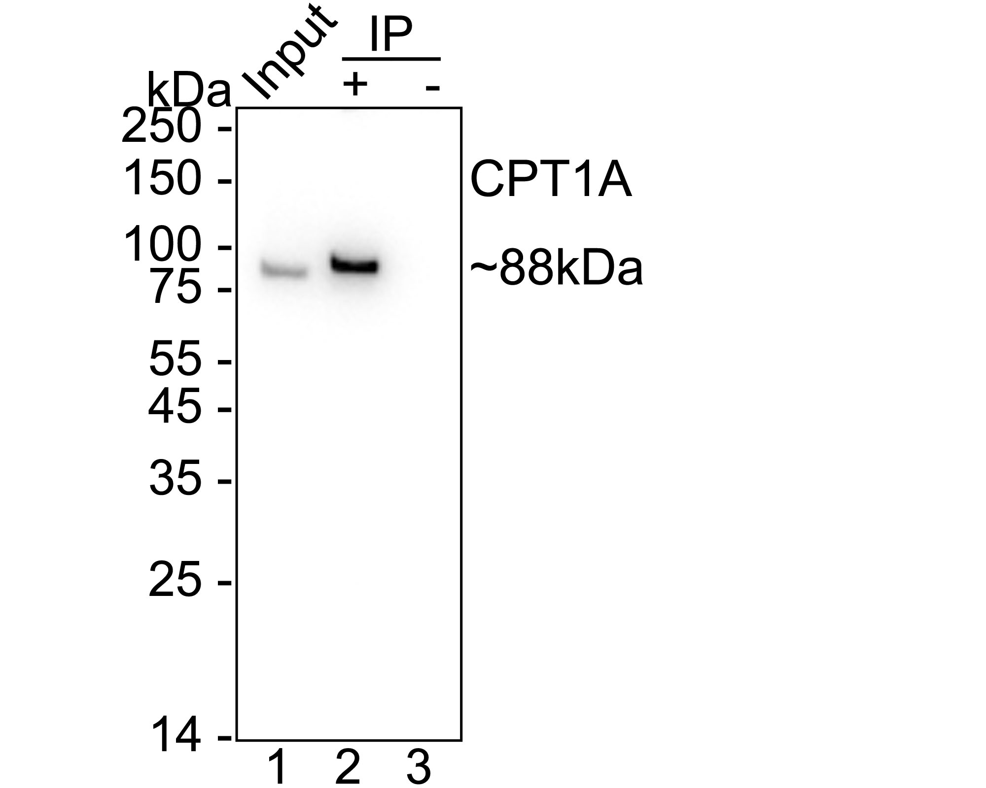
CPT1A was immunoprecipitated from 0.2 mg HeLa cell lysate with Rabbit anti-CPT1A antibody at 2 µg/10 µl beads. Western blot was performed from the immunoprecipitate using Rabbit anti-CPT1A antibody at 1/2,000 dilution. HRP Conjugated Anti-Rabbit IgG for IP secondary antibody at 1/5,000 dilution was used for 1 hour at room temperature. Lane 1: HeLa cell lysate (input), Lane 2:Rabbit anti-CPT1A antibody IP in HeLa cell lysate, Lane 3: Rabbit IgG instead of Rabbit anti-CPT1A antibody in HeLa cell lysate. Blocking/Dilution buffer: 5% NFDM/TBST Exposure time: 4 seconds.| Product Name | CPT1A Recombinant Rabbit Monoclonal Antibody |
|---|---|
| Antibody Type | Primary Antibodies |
| Immunogen | Recombinant protein within human CPT1A aa 201-773 / 773. |
| Clonality | Monoclonal |
|---|---|
| Isotype | IgG |
| Host Species | Rabbit |
| Tested Applications | IHCIPWB |
| WB:1:2000 IHC:1:500-1:2000 IP:1-2μg/sample |
|
| Species Reactivity | HumanMouse |
| Concentration | 1mg/ml |
| Purification | Protein A |
| Gene Symbol | CPT1A |
|---|---|
| Gene Synonyms | CPT1 CPT1-L L-CPT1 |
| Gene Full Name | carnitine palmitoyltransferase 1A |
| Gene Summary | The mitochondrial oxidation of long-chain fatty acids is initiated by the sequential action of carnitine palmitoyltransferase I (which is located in the outer membrane and is detergent-labile) and carnitine palmitoyltransferase II (which is located in the inner membrane and is detergent-stable), together with a carnitine-acylcarnitine translocase. CPT I is the key enzyme in the carnitine-dependent transport across the mitochondrial inner membrane and its deficiency results in a decreased rate of fatty acid beta-oxidation. Alternatively spliced transcript variants encoding different isoforms have been found for this gene. [provided by RefSeq, Jul 2008] |
| Molecular Weight(MW) | 88kDa |
| Cellular Localization | Mitochondrion outer membrane. |
WB
Western blot analysis of CPT1A on different lysates with Rabbit anti-CPT1A antibody at 1/2,000 dilution. Lane 1: 293T cell lysate, Lane 2: HeLa cell lysate, Lane 3: SK-OV-3 cell lysate, Lane 4: MCF7 cell lysate, Lane 5: HepG2 cell lysate, Lane 6: A549 cell lysate, Lane 7: Human kidney tissue lysate, Lysates/proteins at 20 µg/Lane. Exposure time: 30 seconds; 4-20% SDS-PAGE gel. Proteins were transferred to a PVDF membrane and blocked with 5% NFDM/TBST for 1 hour at room temperature. The primary antibody at 1/2,000 dilution was used in 5% NFDM/TBST at 4℃ overnight. Goat Anti-Rabbit IgG - HRP Secondary Antibody at 1/50,000 dilution was used for 1 hour at room temperature.
IHC
Immunohistochemical analysis of paraffin-embedded human kidney tissue with Rabbit anti-CPT1A antibody at 1/2,000 dilution. The section was pre-treated using heat mediated antigen retrieval with Tris-EDTA buffer (pH 9.0) for 20 minutes. The tissues were blocked in 1% BSA for 20 minutes at room temperature, washed with ddH2O and PBS, and then probed with the primary antibody at 1/2,000 dilution for 1 hour at room temperature. The detection was performed using an HRP conjugated compact polymer system. DAB was used as the chromogen. Tissues were counterstained with hematoxylin and mounted with DPX.
IP
CPT1A was immunoprecipitated from 0.2 mg HeLa cell lysate with Rabbit anti-CPT1A antibody at 2 µg/10 µl beads. Western blot was performed from the immunoprecipitate using Rabbit anti-CPT1A antibody at 1/2,000 dilution. HRP Conjugated Anti-Rabbit IgG for IP secondary antibody at 1/5,000 dilution was used for 1 hour at room temperature. Lane 1: HeLa cell lysate (input), Lane 2:Rabbit anti-CPT1A antibody IP in HeLa cell lysate, Lane 3: Rabbit IgG instead of Rabbit anti-CPT1A antibody in HeLa cell lysate. Blocking/Dilution buffer: 5% NFDM/TBST Exposure time: 4 seconds.| Application Notes | WB:1:2000 IHC:1:500-1:2000 IP:1-2μg/sample |
|---|
| Form | Liquid |
|---|---|
| Storage Instructions | Store at +4℃ after thawing. Aliquot store at -20℃. Avoid repeated freeze / thaw cycles. |
| Storage Buffer | PBS (pH7.4), 0.1% BSA, 40% Glycerol. Preservative: 0.05% Sodium Azide. |


 Data sheet for OM643599